. 2006 Apr;80(8):3935-46.
doi: 10.1128/JVI.80.8.3935-3946.2006.

Dissecting rotavirus particle-raft interaction with small interfering RNAs: insights into rotavirus transit through the secretory pathway

Affiliations

Dissecting rotavirus particle-raft interaction with small interfering RNAs: insights into rotavirus transit through the secretory pathway

Mariela A Cuadras et al. J Virol. 2006 Apr.

Abstract

Studies of rotavirus morphogenesis, transport, and release have shown that although these viruses are released from the apical surface of polarized intestinal cells before cellular lysis, they do not follow the classic exocytic pathway. Furthermore, increasing evidence suggests that lipid rafts actively participate in the exit of rotavirus from the infected cell. In this study, we silenced the expression of VP4, VP7, and NSP4 by using small interfering RNAs (siRNAs) and evaluated the effect of shutting down the expression of these proteins on rotavirus-raft interactions. Silencing of VP4 and NSP4 reduced the association of rotavirus particles with rafts; in contrast, inhibition of VP7 synthesis slightly affected the migration of virions into rafts. We found that inhibition of rotavirus migration into lipid rafts, by either siRNAs or tunicamycin, also specifically blocked the targeting of VP4 to rafts, suggesting that the association of VP4 with rafts is mostly mediated by the formation of viral particles in the endoplasmic reticulum (ER). We showed that two populations of VP4 exist, one small population that is independently targeted to rafts and a second large pool of VP4 whose association with rafts is mediated by particle formation in the ER. We also present evidence to support the hypothesis that assembly of VP4 into mature virions takes place in the late stages of transit through the ER. Finally, we analyzed the progression of rotavirus proteins in the exocytic pathway and found that VP4 and virion-assembled VP7 colocalized with ERGIC-53, suggesting that rotavirus particles transit through the intermediate compartment between the ER and the Golgi complex.


Figures
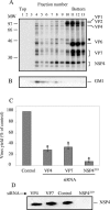
FIG. 1.
FIG. 1.
Isolation of rafts and effects of siRNAs on rotavirus-infected MA104 cells. (A) Pulse-labeled rotavirus proteins associate with rafts in MA104 cells. MA104 cells were infected with DXRRV (MOI of 10) and metabolically labeled as described in Materials and Methods. After lysis in cold 1% TX-100, rafts were purified by equilibrium centrifugation in OptiPrep gradients, fractionated, and analyzed by SDS-PAGE and fluorography. The arrow indicates the migration of the dimeric form of NSP4. (B) Detection of GM1 by Western blotting. Fractions from panel A were subjected to electrophoresis and transferred to PVDF membrane, the membrane was incubated with the biotinylated B subunit of cholera toxin and peroxidase-labeled streptavidin, and a signal was developed by ECL. (C) Effect of siRNAs on rotavirus yield. MA104 cells were transfected with 71.5 pmol of the indicated siRNA, and at 24 h posttransfection, the cells were infected for 14 h with DXRRV at an MOI of 1. The titer of the viral yield was determined on MA104 cells by FFU assay. Titers are expressed as percentages of the virus yield, where 100% represents the titer of virus grown in control transfected cells. Values are means from three experiments, and the standard error is indicated. *, P < 0.05 (one-sample t test). (D) Detection of NSP4 by immunoblotting in the presence of the indicated siRNA. MA104 cells were transfected and infected as described for panel C, and at 12 hpi they were lysed in 1% NP-40 lysis buffer and 10 μg of protein was separated by SDS-PAGE. Electrophoresed proteins were transferred to PVDF membrane, and the membrane was blocked and incubated with MAb B4-2. Bound antibody was detected with peroxidase-labeled anti-mouse IgG and ECL. MW, molecular weight marker (in thousands).
FIG. 2.
FIG. 2.
Rotavirus-raft association is differentially affected by siRNAs. (A) MA104 cells were mock transfected or transfected with the siRNAs that silence the expression of the indicated protein. AT 36 h posttransfection, the cells were infected with DXRRV (MOI, 10) and pulse-labeled as described for Fig. 1A, and rafts were purified by OptiPrep density gradients. After fractionation, 250 μg of 35S-labeled protein of the raft fractions was analyzed by SDS-PAGE and fluorography. For an mβcdx control, 10 mM mβcdx was added to infected cells during starvation and maintained until the end of the experiment. One gel representative of at least three experiments is shown, and the migration of the viral structural proteins and NSP4 is indicated, including the dimeric form of NSP4 (arrow). (B) Densitometric analysis of rotavirus proteins in f4. The radioactive signal for rotavirus proteins VP2, VP4, VP6, VP7, and NSP4 in f4 from gels like the one shown in panel A was quantified with the ChemiImager 4400 low-light imaging system (Alpha Innotech Corp.). Results are expressed as the percentage of these proteins in the raft fraction, where 100% represents the overall amount of these proteins in control transfected cells. (C) Rotavirus infectivity in the raft fraction. Raft fractions from MA104 cells infected with DXRRV and transfected with the indicated siRNAs were normalized to VP6 content by enzyme-linked immunosorbent assay, and virus titers were determined on MA104 cells by FFU assay. Results are expressed as the percentage of infectivity in f4, where 100% represents the infectivity in control lipofected cells. Values are means ± standard errors from at least three experiments. *, P < 0.05 (one-sample t test). Ctrl, control.
FIG. 3.
FIG. 3.
Role of NSP4 in targeting of virions and VP4 to rafts. (A and B) TM treatment prevents the association of rotavirus particles with rafts. Rafts were purified from untreated (A) or TM-treated (B), DXRRV-infected MA104 cells as described in Materials and Methods. OptiPrep density gradients were fractionated and analyzed by SDS-PAGE and fluorography. TM treatment (10 μg/ml) started at 6 hpi and continued to the end of the experiment (B). The positions of viral proteins are indicated at the right, including unglycosylated precursors of VP7 and NSP4 (B) and the dimeric (arrow) form of NSP4 (A). A set of gels representative of at least three experiments is shown. (C) Most of the VP4 associated with rafts comes from viral particles. MA104 cells were infected with DXRRV at an MOI of 10, and at 9 hpi, rafts were purified by cold 1% TX-100 extraction and OptiPrep gradient centrifugation. The rotavirus particles present in the isolated raft fraction were pelleted by ultracentrifugation, and the PUSN was concentrated in a Centriplus YM-10 unit. The presence of VP4 in the pellet (lanes 2 and 5), supernatant (lanes 3 and 6), and semipurified TLPs (lanes 4 and 7) was detected by SDS-PAGE and immunoblotting with MAb HS2. Before ultracentrifugation, aliquots of f4 (lanes 2 and 3) or semipurified TLPs (lane 4) were adjusted to 60 mM octylglucoside, incubated for 30 min at 37°C (lanes 2 to 4), and subjected to ultracentrifugation as described above. Lane 1 represents an aliquot of the starting f4. (D to J) Virion-assembled VP7 colocalizes with PDI in control cells but not in NSP4219 siRNA-transfected cells. MA104 cells grown on coverslips were mock transfected (D to F) or transfected with NSP4219 siRNA (G to J), and at 24 h posttransfection, the cells were infected with DXRRV (MOI, 0.5) for 12 h. The cells were then fixed, permeabilized, and stained with anti-PDI and anti-mouse-FITC. After washing, rotavirus VP7 was detected with MAb 159B (D to H) or 60B (I and J). Bound antibodies were detected with streptavidin-Texas Red. The double-labeled preparations were then incubated with a 1:10,000 dilution of polyclonal rabbit serum (anti-rotavirus R3) and Cy5-labeled anti-rabbit IgG. The coverslips were mounted in glass slides and analyzed in an Eclipse TE300 confocal microscope (Nikon). The left part shows the distribution of PDI in green, and the colocalization with VP7 is shown in yellow. The signal of MAb 159B for panels D and E is shown in panel F. The right part shows the same field but colocalization of rotavirus antigen (R3 stained, blue signal) with VP7 (red signal). All images were obtained with a 60× objective and processed with Adobe Photoshop software. MW, molecular weight marker (in thousands).
FIG. 4.
FIG. 4.
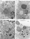
Electron microscopy of MA104 cells transfected with different siRNAs. MA104 cells grown in six-well plates were control transfected or transfected with the indicated siRNA as described in Materials and Methods. At 36 h posttransfection, the cells were infected with DXRRV at an MOI of 10. After 7 h of infection, the cells were recovered, fixed in glutaraldehyde, and processed for transmission electron microscopy. V, viroplasm inclusions; filled arrow, TLPs inside the distended cisternae of the ER; open arrow, DLPs in the cytoplasm; filled arrowhead, enveloped intermediate particle inside the ER; open arrowhead, nonenveloped immature particle inside the ER next to the paracrystalline array.
FIG. 5.
FIG. 5.
Colocalization of ERGIC-53 and rotavirus proteins. MA104 cells grown on coverslips were not infected (A) or infected with DXRRV at an MOI of 0.5 (B to P), and at 12 hpi, cells were fixed and permeabilized and incubated with anti-G1/93 (A to I and P), anti-GPP130 (J to L), and anti-pyruvate dehydrogenase E2/E3bp subunit (M to O) antibodies as markers for the ERGIC, the cis-Golgi, or mitochondria, respectively. After washing, the appropriate secondary antibody was added and then the cells were stained for VP4 with MAb HS2 (C to F) or for virion-assembled VP7 with MAb 159B (G to P) as described in Materials and Methods. The nucleus was costained with a 1:20,000 dilution of TOTO-3, and the preparations were mounted and analyzed by confocal microscopy as described in the legend to Fig. 3. In all images, the green signal shows the distribution of a cellular organelle and the red signal shows the distribution of rotavirus proteins. (P) Representative sequential sections with a thickness of 1 μm acquired from the bottom to the top of panel I. All images were obtained with a 60× objective; panels A, B, and F show a 2× zoom image. The digital images were processed with Adobe Photoshop.

Similar articles

Cited by

References

    1. Aridor, M., S. I. Bannykh, T. Rowe, and W. E. Balch. 1995. Sequential coupling between COPII and COPI vesicle coats in endoplasmic reticulum to Golgi transport. J. Cell Biol. 131:875-893. - PMC - PubMed
    1. Bannykh, S. I., T. Rowe, and W. E. Balch. 1996. The organization of endoplasmic reticulum export complexes. J. Cell Biol. 135:19-35. - PMC - PubMed
    1. Ben-Tekaya, H., K. Miura, R. Pepperkok, and H. P. Hauri. 2005. Live imaging of bidirectional traffic from the ERGIC. J. Cell Sci. 118:357-367. - PubMed
    1. Brown, D. A., and J. K. Rose. 1992. Sorting of GPI-anchored proteins to glycolipid-enriched membrane subdomains during transport to the apical cell surface. Cell 68:533-544. - PubMed
    1. Ciarlet, M., S. E. Crawford, and M. K. Estes. 2001. Differential infection of polarized epithelial cell lines by sialic acid-dependent and sialic acid-independent rotavirus strains. J. Virol. 75:11834-11850. - PMC - PubMed

Publication types

MeSH terms

LinkOut - more resources