. 2006 Apr;80(8):4005-16.
doi: 10.1128/JVI.80.8.4005-4016.2006.

The products of the herpes simplex virus type 1 immediate-early US1/US1.5 genes downregulate levels of S-phase-specific cyclins and facilitate virus replication in S-phase Vero cells

Affiliations

The products of the herpes simplex virus type 1 immediate-early US1/US1.5 genes downregulate levels of S-phase-specific cyclins and facilitate virus replication in S-phase Vero cells

Joseph S Orlando et al. J Virol. 2006 Apr.

Abstract

Herpes simplex virus type 1 ICP22-/U(S)1.5- mutants initiate viral gene expression in all cells; however, in most cell types, the replication process stalls due to an inability to express gamma2 late proteins. Although the function of ICP22/U(S)1.5 has not been established, it has been suggested that these proteins activate, induce, or repress the activity of cellular proteins during infection. In this study, we hypothesized that cell cycle-associated proteins are targets of ICP22/U(S)1.5. For this purpose, we first isolated and characterized an ICP22-/U(S)1.5- mutant virus, 22/n199. Like other ICP22-/U(S)1.5- mutants, 22/n199 replicates in a cell-type-specific manner and fails to induce efficient gamma2 late gene expression in restrictive cells. Although synchronization of restrictive human embryonic lung cells in each phase of the cell cycle did not overcome the growth restrictions of 22/n199, synchronization of permissive Vero cells in S phase rendered them less able to support 22/n199 plaque formation and replication. Consistent with this finding, expression of cellular S-phase cyclins was altered in an ICP22/U(S)1.5-dependent manner specifically when S-phase Vero cells were infected. Collectively, these observations support the notion that ICP22/U(S)1.5 deregulates the cell cycle upon infection of S-phase permissive cells by altering expression of key cell cycle regulatory proteins either directly or indirectly.


Figures
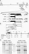
FIG. 1.
FIG. 1.
Southern blot analysis of wild-type, 22/n199, and 22/n199R genomes. (A) Diagram of the 152-kb HSV-1 genome and existing ICP22/US1.5 mutants. The HSV-1 genome is composed of the unique long (UL), unique short (US), internal (IR), and terminal (TR) repeats. Depicted above and below the genome are the mRNAs for the five IE genes and the L/ST transcripts. The 3.3-kb EcoRI-KpnI fragment containing the US1 gene is expanded. The open reading frames for ICP22, US1.5, and US2 are shown in black, gray, and white boxes, respectively. Two existing ICP22/US1.5 mutants as well as the 22/n199 mutant isolated in this study are illustrated. Deleted sequences are not illustrated. The hatched box shows the LacZ replacement mutation. Sequences after the inserted stop codon of 22/n199 are illustrated by open boxes. (B) The 15.1-kb EcoRI H fragment of the HSV-1 genome, encompassing all of the US region and portions of two repeats (24). Positions and sizes of relevant DNA fragments (lines A to E) and positions of restriction enzyme sites within the genomes, as well as the ICP22 and US1.5 open reading frames and transcripts, are illustrated. (C) Southern blot. Vero cells were infected with two viral stocks each of the wild-type virus, 22/n199, and 22/n199R. Viral DNA was harvested and 1 μg of DNA was digested with EcoRI and HpaI (left) or EcoRI and PvuII (right). Viral DNA was separated by agarose gel electrophoresis and analyzed by Southern blot hybridization using the 1.2-kb probe encompassing the entire ICP22 open reading frame indicated in panel B. The scale between lane sets represents kilobase pairs.
FIG. 2.
FIG. 2.
Growth properties of the wild-type virus, 22/n199, and 22/n199R in Vero, RAB-9, HEL, and AGMK cells. (A) One-step replication assay. Replicate plates of Vero, RAB-9, HEL, and AGMK cells were infected with the wild-type virus, 22/n199, or 22/n199R at an MOI of 2.5 PFU/cell. Cells were harvested at the indicated times postinfection, and the amount of virus was determined by plaque assay on Vero cell monolayers. Each point represents the average of results of three independent infections. (B) Viral DNA slot blot. Vero, RAB-9, HEL, and AGMK cells were infected with the wild-type virus or 22/n199 at an MOI of 5 PFU/cell. Cells were harvested at the indicated times, total DNA was isolated, and 5 μg of DNA was transferred to a nylon membrane. Viral DNA was detected following hybridization to a cDNA probe generated by random priming in the presence of [α-P32]dCTP of EcoRI A, D, I, N, and O fragments of the HSV-1 genome. (C) Plaque size. Concentrated viral stocks of the wild-type virus, 22/n199, and 22/n199R were serially diluted and used to infect Vero or RAB-9 cells. Following infection, cells were overlaid with methylcellulose-containing medium for 4 days. Cells were fixed and stained with 0.1% crystal violet in 20% ethanol. Photographs of representative plaques are shown. (D) Expression of ICP22 and γ1 and γ2 L proteins in Vero and RAB-9 cells. Actively dividing Vero and RAB-9 cells were seeded on 35-mm petri dishes. After 24 h, cells were infected with either the wild-type virus (wt) or 22/n199 (9) at an MOI of 2.5 PFU/cell. At the time of infection and every 3 h thereafter, cells were harvested and lysed, volumes were standardized for cell equivalents, and separation by SDS-PAGE and Western blots for ICP22, gE (γ1), and US11 (γ2) were performed.
FIG. 3.
FIG. 3.
Plating efficiency and replication of the wild-type virus and 22/n199 on restrictive and permissive cells synchronized by isoleucine deprivation and double thymidine blocks. Actively dividing HEL (A to C) or Vero (D to I) cells were seeded on 60-mm petri dishes. After 24 h, cells were blocked in either early S by double thymidine block or in G1 by isoleucine deprivation. Following cell cycle arrest, all cells were released into normal medium and examined every 3 h for 24 h for cell cycle analysis or plating efficiency. (A, D, and G) Cell cycle analysis. Cells and medium were collected and fixed in 70% ethanol. DNA content was determined by treating cells with 0.005% propidium iodide and 0.004% RNase and monitoring fluorescence by FACS. Each point represents an average of results for three independent plates. (B, E, and H) Plating efficiency. Plates were infected with ∼50 PFU (B) or ∼100 PFU (E and H) of the wild-type virus or 22/n199 viruses and overlaid with normal medium containing 0.5% methylcellulose. Four days postinfection, cells were fixed and stained with 0.1% crystal violet in 20% ethanol. Each point represents an average of results for three independent infections. (C, F, and I) Virus replication. Cells synchronized to each phase of the cell cycle and asynchronous cells were infected with 2.5 PFU/cell of the wild-type virus or 22/n199. HEL cells synchronized by double thymidine block were harvested at 12, 6, and 9 h post-release of the double thymidine block to represent G1, S, and G2/M cells, respectively. Vero cells synchronized by isoleucine deprivation were infected at 9, 15, and 18.5 h postrelease of the G1 block and represent G1, S, and G2/M phase cells, respectively. Vero cells synchronized by double thymidine block were infected at 15, 3, and 9 h postrelease for G1, S, and G2/M cells, respectively. At 24 hpi, cells were harvested and the amounts of virus present determined by plaque assay on Vero cells. Each bar represents the average of results for three independent infections. Numbers above the bars representing 22/n199 replication are the output titers in restrictive cells. Error bars represent the standard deviations. These results are representative of at least three independent experiments.
FIG. 4.
FIG. 4.
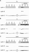
ICP22 and/or US1.5 protein expression affects levels of S- and G2-specific cyclins during infection. Vero cells synchronized in G1 (A), S (B), and G2/M (C) by isoleucine deprivation and release were mock infected (lane M) or infected with 10 PFU/cell of the wild type (lane wt) or 22/n199 (lane 9) in the absence (left) and presence (right) of the proteasome inhibitor MG132 (2.5 μM). At the time of infection, cells were also collected for analysis (lane 0). Twelve hours postinfection, cells were harvested and lysed, volume was standardized for cell equivalents, and duplicate samples of proteins were separated by SDS-PAGE. Proteins were transferred to nitrocellulose, and Western blots of cyclin A, cyclin B, and cyclin E were performed.

Similar articles

Cited by

References

    1. Ackermann, M., M. Sarmiento, and B. Roizman. 1985. Application of antibody to synthetic peptides for characterization of the intact and truncated α22 protein specified by herpes simplex virus 1 and the R325 α22− deletion mutant. J. Virol. 56:207-215. - PMC - PubMed
    1. Advani, S. J., R. Brandimarti, R. R. Weichselbaum, and B. Roizman. 2000. The disappearance of cyclins A and B and the increase in activity of the G2/M-phase cellular kinase cdc2 in herpes simplex virus 1-infected cells require expression of the α22/US1.5 and UL13 viral genes. J. Virol. 74:8-15. - PMC - PubMed
    1. Advani, S. J., R. R. Weichselbaum, and B. Roizman. 2000. The role of cdc2 in the expression of herpes simplex virus genes. Proc. Natl. Acad. Sci. USA 97:10996-11001. - PMC - PubMed
    1. Advani, S. J., R. R. Weichselbaum, and B. Roizman. 2003. Herpes simplex virus 1 activates cdc2 to recruit topoisomerase II alpha for post-DNA synthesis expression of late genes. Proc. Natl. Acad. Sci. USA 100:4825-4830. - PMC - PubMed
    1. Aleem, E., H. Kiyokawa, and P. Kaldis. 2005. Cdc2-cyclin E complexes regulate the G1/S phase transition. Nat. Cell Biol. 7:831-836. - PubMed

LinkOut - more resources