Mucosal immunization with surface-displayed severe acute respiratory syndrome coronavirus spike protein on Lactobacillus casei induces neutralizing antibodies in mice
- PMID: 16571824
- PMCID: PMC1440448
- DOI: 10.1128/JVI.80.8.4079-4087.2006
Mucosal immunization with surface-displayed severe acute respiratory syndrome coronavirus spike protein on Lactobacillus casei induces neutralizing antibodies in mice
Abstract
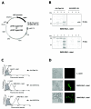
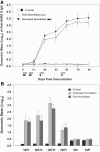
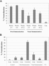
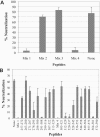
Induction of mucosal immunity may be important for preventing SARS-CoV infections. For safe and effective delivery of viral antigens to the mucosal immune system, we have developed a novel surface antigen display system for lactic acid bacteria using the poly-gamma-glutamic acid synthetase A protein (PgsA) of Bacillus subtilis as an anchoring matrix. Recombinant fusion proteins comprised of PgsA and the Spike (S) protein segments SA (residues 2 to 114) and SB (residues 264 to 596) were stably expressed in Lactobacillus casei. Surface localization of the fusion protein was verified by cellular fractionation analyses, immunofluorescence microscopy, and flow cytometry. Oral and nasal inoculations of recombinant L. casei into mice resulted in high levels of serum immunoglobulin G (IgG) and mucosal IgA, as demonstrated by enzyme-linked immunosorbent assays using S protein peptides. More importantly, these antibodies exhibited potent neutralizing activities against severe acute respiratory syndrome (SARS) pseudoviruses. Orally immunized mice mounted a greater neutralizing-antibody response than those immunized intranasally. Three new neutralizing epitopes were identified on the S protein using a peptide neutralization interference assay (residues 291 to 308, 520 to 529, and 564 to 581). These results indicate that mucosal immunization with recombinant L. casei expressing SARS-associated coronavirus S protein on its surface provides an effective means for eliciting protective immune response against the virus.
Figures


Similar articles
-
A single immunization with a rhabdovirus-based vector expressing severe acute respiratory syndrome coronavirus (SARS-CoV) S protein results in the production of high levels of SARS-CoV-neutralizing antibodies.J Gen Virol. 2005 May;86(Pt 5):1435-1440. doi: 10.1099/vir.0.80844-0. J Gen Virol. 2005. PMID: 15831955 Free PMC article.
-
Surface-displayed porcine epidemic diarrhea viral (PEDV) antigens on lactic acid bacteria.Vaccine. 2007 Dec 21;26(1):24-31. doi: 10.1016/j.vaccine.2007.10.065. Epub 2007 Nov 20. Vaccine. 2007. PMID: 18054413 Free PMC article.
-
SARS coronavirus spike polypeptide DNA vaccine priming with recombinant spike polypeptide from Escherichia coli as booster induces high titer of neutralizing antibody against SARS coronavirus.Vaccine. 2005 Oct 10;23(42):4959-68. doi: 10.1016/j.vaccine.2005.05.023. Vaccine. 2005. PMID: 15993989 Free PMC article.
-
SARS vaccine development.Emerg Infect Dis. 2005 Jul;11(7):1016-20. doi: 10.3201/1107.050219. Emerg Infect Dis. 2005. PMID: 16022774 Free PMC article. Review.
-
Severe acute respiratory syndrome: vaccine on the way.Chin Med J (Engl). 2005 Sep 5;118(17):1468-76. Chin Med J (Engl). 2005. PMID: 16157050 Review. No abstract available.
Cited by
-
Display of recombinant proteins at the surface of lactic acid bacteria: strategies and applications.Microb Cell Fact. 2016 May 3;15:70. doi: 10.1186/s12934-016-0468-9. Microb Cell Fact. 2016. PMID: 27142045 Free PMC article. Review.
-
Sublingual immunization with recombinant adenovirus encoding SARS-CoV spike protein induces systemic and mucosal immunity without redirection of the virus to the brain.Virol J. 2012 Sep 21;9:215. doi: 10.1186/1743-422X-9-215. Virol J. 2012. PMID: 22995185 Free PMC article.
-
Probiotic-Based Vaccines May Provide Effective Protection against COVID-19 Acute Respiratory Disease.Vaccines (Basel). 2021 May 6;9(5):466. doi: 10.3390/vaccines9050466. Vaccines (Basel). 2021. PMID: 34066443 Free PMC article. Review.
-
Comparison of the immune responses induced by oral immunization of mice with Lactobacillus casei-expressing porcine parvovirus VP2 and VP2 fused to Escherichia coli heat-labile enterotoxin B subunit protein.Comp Immunol Microbiol Infect Dis. 2011 Jan;34(1):73-81. doi: 10.1016/j.cimid.2010.02.004. Epub 2010 Mar 11. Comp Immunol Microbiol Infect Dis. 2011. PMID: 20226529 Free PMC article.
-
Synthetic reconstruction of zoonotic and early human severe acute respiratory syndrome coronavirus isolates that produce fatal disease in aged mice.J Virol. 2007 Jul;81(14):7410-23. doi: 10.1128/JVI.00505-07. Epub 2007 May 16. J Virol. 2007. PMID: 17507479 Free PMC article.
References
-
- Ashiuchi, M., C. Nawa., T. Kamei, J. J. Song, S. P. Hong, M. H. Sung, K. Soda, T. Yagi, and H. Misono. 2001. Physiological and biochemical characteristics of poly-γ-glutamate synthetase complex of Bacillus subtilis. Eur. J. Biochem. 268:5321-5328. - PubMed
-
- Ashiuchi, M., K. Soda, and H. Misono. 1999. A poly-γ-glutamate synthetic system of Bacillus subtilis IFO 3336: gene cloning and biochemical analysis of poly-γ-glutamate produced by Escherichia coli clone cells. Biochem. Biophys. Res. Commun. 263:6-12. - PubMed
-
- Bukreyev, A., E. W. Lamirande, U. J. Buchholz, L. N. Vogel, W. R. Elkins, M. St. Claire, B. R. Murphy, K. Subbarao, and P. L. Collins. 2004. Mucosal immunization of African green monkeys (Cercopithecus aethlops) with an attenuated parainfluenza virus expressing the SARS coronavirus spike protein for the prevention of SARS. Lancet 363:2122-2127. - PMC - PubMed
Publication types
MeSH terms
Substances
Grants and funding
LinkOut - more resources
Full Text Sources
Other Literature Sources
Miscellaneous

