Recovery of a recombinant salmonid alphavirus fully attenuated and protective for rainbow trout
- PMID: 16571825
- PMCID: PMC1440445
- DOI: 10.1128/JVI.80.8.4088-4098.2006
Recovery of a recombinant salmonid alphavirus fully attenuated and protective for rainbow trout
Abstract
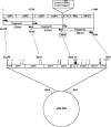
Sleeping disease virus (SDV) is a member of the new Salmonid alphavirus genus within the Togaviridae family. The single-stranded RNA genome of SDV is 11,894 nucleotides long, excluding the 3' poly(A) tail. A full-length cDNA has been generated; the cDNA was fused to a hammerhead ribozyme sequence at the 5' end and inserted into a transcription plasmid (pcDNA3) backbone, yielding pSDV. By transfection of pSDV into fish cells, recombinant SDV (rSDV) was successfully recovered. Demonstration of the recovery of rSDV was provided by immunofluorescence assay on rSDV-infected cells and by the presence of a genetic tag, a BlpI restriction enzyme site, introduced into the rSDV RNA genome. SDV infectious cDNA was used for two kinds of experiments (i) to evaluate the impact of various targeted mutations in nsP2 on viral replication and (ii) to study the virulence of rSDV in trout. For the latter aspect, when juvenile trout were infected by immersion in a water bath with the wild-type virus-like rSDV, no deaths or signs of disease appeared in fish, although they were readily infected. In contrast, cumulative mortality reached 80% in fish infected with the wild-type SDV (wtSDV). When rSDV-infected fish were challenged with wtSDV 3 and 5 months postinfection, a long-lasting protection was demonstrated. Interestingly, a variant rSDV (rSDV14) adapted to grow at a higher temperature, 14 degrees C instead of 10 degrees C, was shown to become pathogenic for trout. Comparison of the nucleotide sequences of wtSDV, rSDV, and rSDV14 genomes evidenced several amino acid changes, and some changes may be linked to the pathogenicity of SDV in trout.
Figures


Similar articles
-
A fully attenuated recombinant Salmonid alphavirus becomes pathogenic through a single amino acid change in the E2 glycoprotein.J Virol. 2013 May;87(10):6027-30. doi: 10.1128/JVI.03501-12. Epub 2013 Feb 28. J Virol. 2013. PMID: 23449806 Free PMC article.
-
Rainbow trout sleeping disease virus is an atypical alphavirus.J Virol. 2000 Jan;74(1):173-83. doi: 10.1128/jvi.74.1.173-183.2000. J Virol. 2000. PMID: 10590104 Free PMC article.
-
Comparison of two aquatic alphaviruses, salmon pancreas disease virus and sleeping disease virus, by using genome sequence analysis, monoclonal reactivity, and cross-infection.J Virol. 2002 Jun;76(12):6155-63. doi: 10.1128/jvi.76.12.6155-6163.2002. J Virol. 2002. PMID: 12021349 Free PMC article.
-
Alphavirus infections in salmonids--a review.J Fish Dis. 2007 Sep;30(9):511-31. doi: 10.1111/j.1365-2761.2007.00848.x. J Fish Dis. 2007. PMID: 17718707 Review.
-
Reverse genetics on fish rhabdoviruses: tools to study the pathogenesis of fish rhabdoviruses.Curr Top Microbiol Immunol. 2005;292:119-41. doi: 10.1007/3-540-27485-5_6. Curr Top Microbiol Immunol. 2005. PMID: 15981470 Review.
Cited by
-
A fully attenuated recombinant Salmonid alphavirus becomes pathogenic through a single amino acid change in the E2 glycoprotein.J Virol. 2013 May;87(10):6027-30. doi: 10.1128/JVI.03501-12. Epub 2013 Feb 28. J Virol. 2013. PMID: 23449806 Free PMC article.
-
A Viral mRNA Motif at the 3'-Untranslated Region that Confers Translatability in a Cell-Specific Manner. Implications for Virus Evolution.Sci Rep. 2016 Jan 12;6:19217. doi: 10.1038/srep19217. Sci Rep. 2016. PMID: 26755446 Free PMC article.
-
The reverse genetics applied to fish RNA viruses.Vet Res. 2011 Jan 24;42(1):12. doi: 10.1186/1297-9716-42-12. Vet Res. 2011. PMID: 21314978 Free PMC article. Review.
-
A 6K-deletion variant of salmonid alphavirus is non-viable but can be rescued through RNA recombination.PLoS One. 2014 Jul 10;9(7):e100184. doi: 10.1371/journal.pone.0100184. eCollection 2014. PLoS One. 2014. PMID: 25009976 Free PMC article.
-
Viruses of fish: an overview of significant pathogens.Viruses. 2011 Nov;3(11):2025-46. doi: 10.3390/v3112025. Epub 2011 Oct 25. Viruses. 2011. PMID: 22163333 Free PMC article. Review.
References
-
- Anonymous. 1985. Maladie du sommeil et pollutions expérimentales. Technical report no. 118. Laboratoire National de Pathologie des Animaux Aquatiques, CNEVA, Brest, France.
-
- Boucher, P., J. Castric, and F. Baudin Laurencin. 1994. Observation of virus-like particles in rainbow-trout Oncorhynchus mykiss infected with sleeping disease virulent material. Bull. Eur. Assoc. Fish Pathol. 14:215-216.
-
- Boucher, P. 1995. Ph.D. thesis. Université de Rennes I, Rennes, France.
-
- Castric, J., F. Baudin Laurencin, M. Brémont, J. Jeffroy, A. Le Ven, and M. Bearzotti. 1997. Isolation of the virus responsible for sleeping disease in experimentally infected rainbow trout (Oncorhynchus mykiss). Bull. Eur. Assoc. Fish Pathol. 17:27-30.
-
- Davis, N. L., L. V. Willis, J. F. Smith, and R. E. Johnston. 1989. In vitro synthesis of infectious Venezuelan equine encephalitis virus RNA from a cDNA clone: analysis of a viable deletion mutant. Virology 171:189-204. - PubMed
Publication types
MeSH terms
LinkOut - more resources
Full Text Sources
Other Literature Sources

