. 2006 Sep;291(3):C483-9.
doi: 10.1152/ajpcell.00097.2006. Epub 2006 Mar 29.

Role of vimentin in smooth muscle force development

Affiliations

Role of vimentin in smooth muscle force development

Ruping Wang et al. Am J Physiol Cell Physiol. 2006 Sep.

Abstract

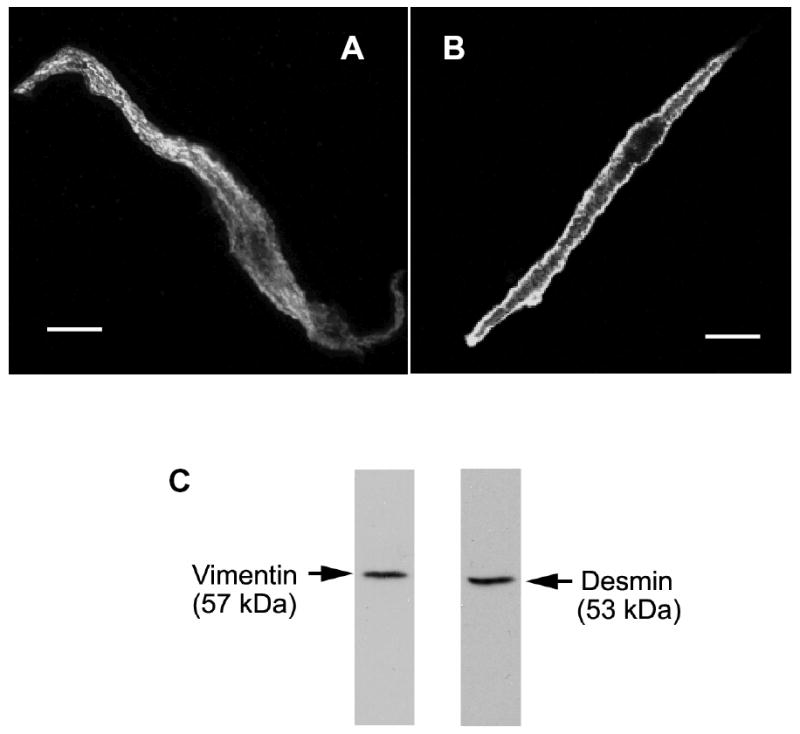
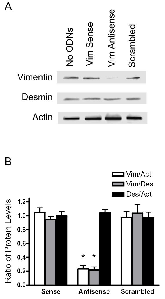
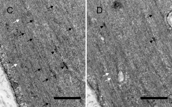
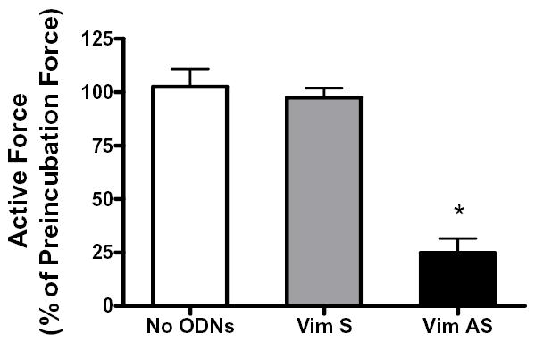
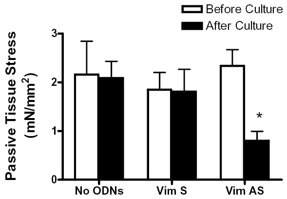
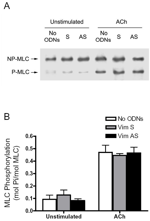
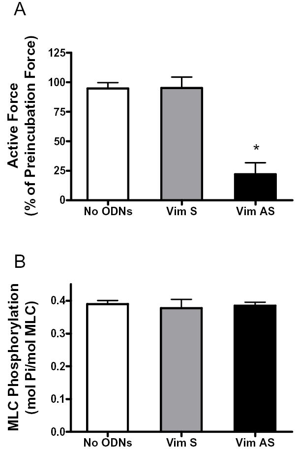
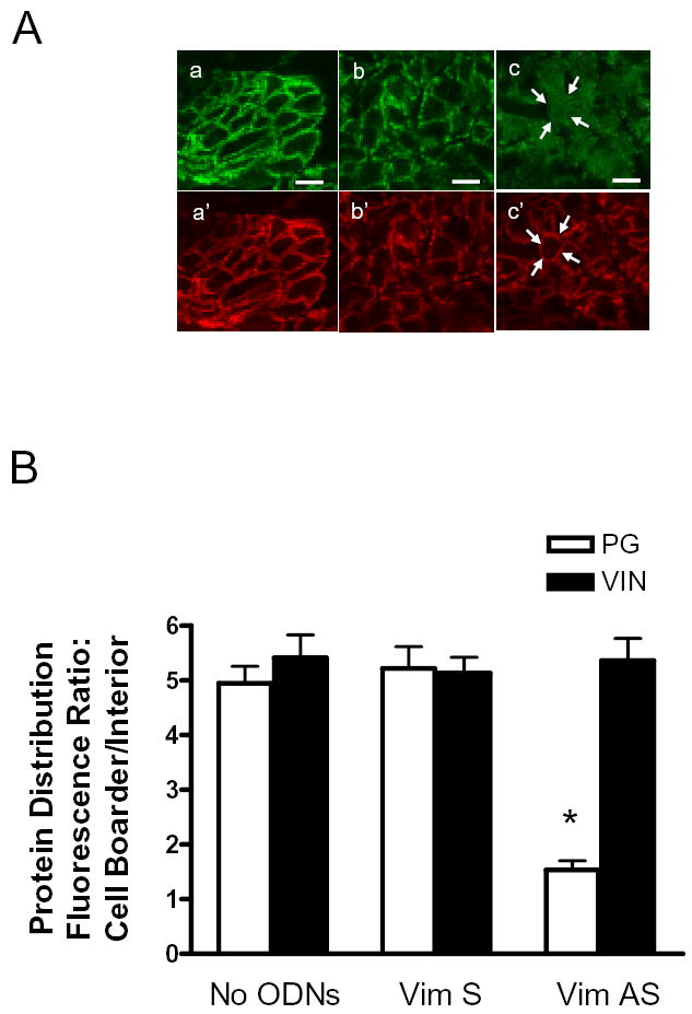
Vimentin intermediate filaments undergo spatial reorganization in cultured smooth muscle cells in response to contractile activation; however, the role of vimentin in the physiological properties of smooth muscle has not been well elucidated. Tracheal smooth muscle strips were loaded with antisense oligonucleotides (ODNs) against vimentin and then cultured for 2 days to allow for protein degradation. Treatment with vimentin antisense, but not sense, ODNs suppressed vimentin protein expression; neither vimentin antisense nor sense ODNs affected protein levels of desmin and actin. Force development in response to ACh stimulation or KCl depolarization was lower in vimentin-deficient tissues than in vimentin sense ODN- or non-ODN-treated muscle strips. Passive tension was also depressed in vimentin-depleted muscle tissues. Vimentin downregulation did not attenuate increases in myosin light chain (MLC) phosphorylation in response to contractile stimulation or basal MLC phosphorylation. In vimentin sense ODN-treated or non-ODN-treated smooth muscle strips, the desmosomal protein plakoglobin was primarily localized in the cell periphery. The membrane-associated localization of plakoglobin was reduced in vimentin-depleted muscle tissues. These studies suggest that vimentin filaments play an important role in mediating active force development and passive tension, which are not regulated by MLC phosphorylation. Vimentin downregulation impairs the structural organization of desmosomes, which may be associated with the decrease in force development.


Figures

Figure 1
Figure 1. Subcellular distribution of vimentin and desmin in freshly-dissociated smooth muscle cells
Smooth muscle cells were freshly dissociated from canine tracheal smooth muscle tissues. These cells were then immunofluorescently labeled for vimentin (A) or for desmin (B). Vimentin is distributed throughout the myoplasm, whereas desmin is more concentrated at the membrane. The nucleus appeared as a dark area. Bar, 5 μm. C, immunoblots of muscle extracts were obtained with antibodies against vimentin or desmin.
Figure 2
Figure 2. Downregulation of vimentin protein by vimentin antisense oligodeoxynucleotides (ODNs)
A, blots of protein extracts from muscle strips that had been cultured for 2 days without ODNs (No ODNs), with vimentin (Vim) sense ODNs or vimentin antisense ODNs, or scrambled ODNs were probed with antibodies against vimentin, desmin, and actin. The level of vimentin was lower in antisense-treated muscle strips than in strips not treated with ODNs, or sense-treated or scrambled sequence-treated muscle strips. Similar amounts of desmin and actin were detected in all four strips. Molecular mass markers are indicated on left. B, ratios of protein expression obtained from muscle strips treated with vimentin sense or antisense, or scrambled ODNs. Ratios in sense-treated, antisense-treated or scrambled sequence-treated muscle strips are normalized to ratios obtained in muscle strips not treated with ODNs. Vim, vimentin; Act, actin; Des, desmin. Values represent mean ± SE (n = 4–6). Asterisk indicates significantly lower ratios in antisense-treated strips as compared to no-ODN-treated, sense-treated and scrambled sequence-treated muscle strips (P < 0.05). C & D, representative electron micrographs of the longitudinal sections of smooth muscle tissues treated with vimentin sense (C) and tissues treated with antisense ODNs (D). Black arrows indicate intermediate filaments; white arrows, thin filaments; black arrow heads, thick filaments. Bar, 200 nm.
Figure 3
Figure 3. Effects of vimentin depletion on contractile force in response to stimulation with acetylcholine
The contractile responses of canine tracheal smooth muscle strips were assessed, after which vimentin sense or vimentin antisense was loaded into these muscle strips, and these strips were incubated for 2 days to allow for protein depletion. The contractile responses of these muscle strips were then determined. Vimentin antisense inhibited ACh-induced contraction in smooth muscle strips after 2-day incubation. The contractile responses of no-ODN-treated or sense-treated muscle strips were similar to preincubation force after 2 days of culture. Mean active force in response to 10−5M ACh was quantitated as percent of ACh-induced force in each strip before incubation. Values are mean ± SE. Asterisk indicates significantly lower response as compared to muscles treated with sense ODNs or not treated with ODNs (n = 10, P < 0.05).
Figure 4
Figure 4. Passive tension in smooth muscle strips treated with vimentin antisense
The passive tension of tracheal smooth muscle strips that had been treated with vimentin sense or antisense, or not treated with ODNs was determined before and after the organ culture. The passive tension in muscle strips treated with sense or in strips not treated with ODNs was comparable to preincubation tension after 2-day culture. The tension in muscle strips exposed to vimentin antisense was decreased after the incubation. Values are mean ± SE. Asterisk indicates significantly lower tension as compared to the value before incubation (n = 10, P < 0.05).
Figure 5
Figure 5. Effects of vimentin downregulation on myosin light chain phosphorylation
A, representative immunoblot showing unstimulated or ACh (10−5M, 5 min) stimulated myosin light chain (MLC) phosphorylation in tracheal smooth muscle strips that had been incubated with no ODNs, or with vimentin sense ODNs (S) or vimentin antisense ODNs (AS). NP-MLC, non-phosphorylated MLC; P-MLC, phosphorylated MLC. B, Differences between antisense-treated, sense-treated, and no ODNs-treated strips were not statistically significant (P > 0.05). Values shown are mean ± SE (n = 4–5).
Figure 6
Figure 6. Contractile force and myosin light chain phosphorylation in vimentin-deficient muscle strips stimulated by KCl
A, smooth muscle strips were contracted with 60 mM KCl before and after 2-day incubation without ODNs, or with vimentin sense or antisense. Mean active force in response to 60 mM KCl was quantitated as percent of KCl-induced force in each strip before incubation. Values are mean ± SE. Asterisk indicates significantly lower response as compared to muscles not treated with ODNs, or treated with sense ODNs (n = 6–7) (P < 0.05). B, myosin light chain phosphorylation in muscle strips incubated without ODNs, or with vimentin sense or antisense. Myosin light chain phosphorylation was measured in smooth muscle strips 5 min after stimulation with 60 mM KCl. Differences between antisense-treated, sense-treated and no ODNs-treated strips were not statistically significant. Values shown are mean ± SE (n = 4).
Figure 7
Figure 7. Subcellular distribution of plakoglobin in vimentin-depleted smooth muscle tissues
Tracheal smooth muscle strips that had been treated with vimentin antisense or sense oligonucleotides, or without oligonucleotides were cryosectioned. These sections were then immunofluorescently labeled for plakoglobin and vinculin. A, plakoglobin is mainly localized on the membrane in cells not treated with oligonucelotides (a) and in cells treated with vimentin sense oligonucleotides (b). The membrane distribution of plakoglobin is reduced in tissues treated with vimentin antisense oligonucleotides (c). However, vinculin localization is similar in tissues not treated with oligonucleotides (a’) and in muscle strips treated with vimentin sense (b’) or antisense (c’) oligonuceleotides. Bar, 5 μm. Arrows indicate the same cross section of the cell in two different images (c and c’). B, mean ratios of fluorescence intensity for plakoglobin (PG) and vinculin (VIN) in tracheal smooth muscle tissues not treated with ODNs (no ODNs), or treated with vimentin sense (Vim S) or vimentin antisense (Vim AS) oligonucleotides. Protein distribution in cells of the tissue cross-sections was expressed as the ratio of pixel intensity of the cell boarder to the cell interior (See Materials and methods). Each mean value was obtained from an average of 3–4 line scans in each of 20 no-ODN-treated cells, 20 sense-treated cells, and 20 antisense-treated cells from 3 experiments. * The fluorescence intensity ratio for plakoglobin in the antisense-treated muscles was significantly lower as compared to muscle tissues not treated with ODNs or tissues treated with sense ODNs (P < 0.05).

Similar articles

Cited by

References

    1. Ashton FT, Somlyo AV, Somlyo AP. The contractile apparatus of vascular smooth muscle: intermediate high voltage stereo electron microscopy. J Mol Biol. 1975;98:17–29. - PubMed
    1. Barany M, Barron JT, Gu L, Barany K. Exchange of the actin-bound nucleotide in intact arterial smooth muscle. J Biol Chem. 2001;276:48398–48403. - PubMed
    1. Bornslaeger EA, Godsel LM, Corcoran CM, Park JK, Hatzfeld M, Kowalczyk AP, Green KJ. Plakophilin 1 interferes with plakoglobin binding to desmoplakin, yet together with plakoglobin promotes clustering of desmosomal plaque complexes at cell-cell borders. J Cell Sci. 2001;114:727–738. - PubMed
    1. Chan W, Kozma R, Yasui Y, Inagaki M, Leung T, Manser E, Lim L. Vimentin intermediate filament reorganization by Cdc42: involvement of PAK and p70 S6 kinase. Eur J Cell Biol. 2002;81:692–701. - PubMed
    1. Chang L, Goldman RD. Intermediate filaments mediate cytoskeletal crosstalk. Nat Rev Mol Cell Biol. 2004;5:601–613. - PubMed

Publication types

MeSH terms

LinkOut - more resources