. 2006;13(1):27-40.
doi: 10.3727/000000006783991917.

Molecular cloning of the m-Golsyn gene and its expression in the mouse brain

Affiliations

Molecular cloning of the m-Golsyn gene and its expression in the mouse brain

Eishi Funakoshi et al. Gene Expr. 2006.

Abstract

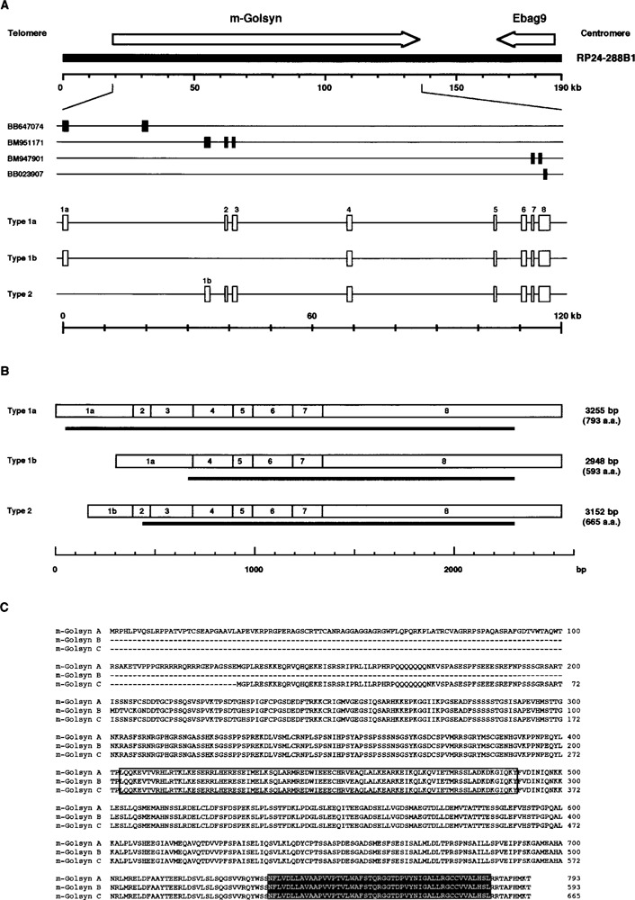
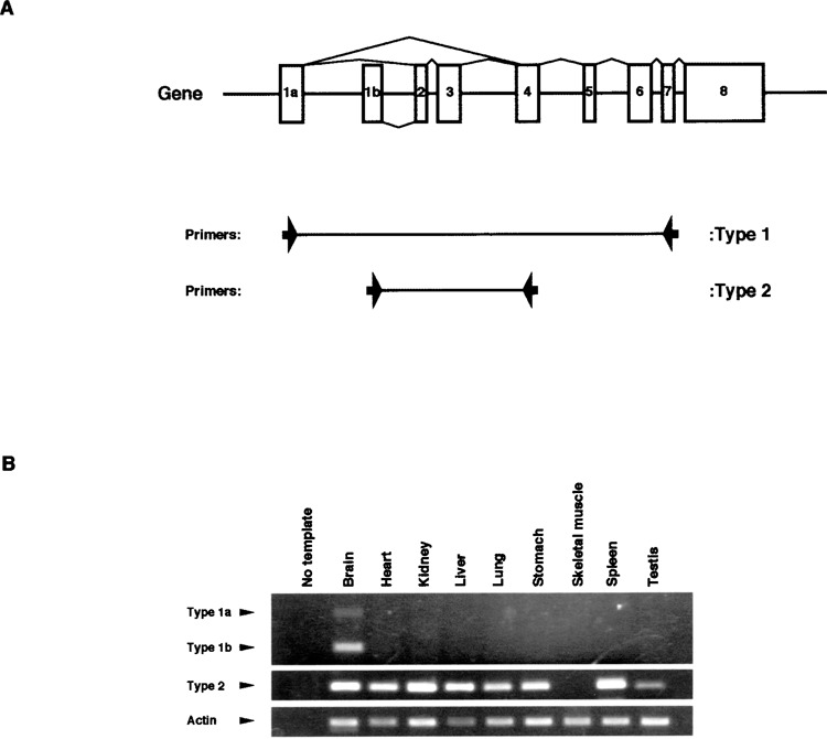
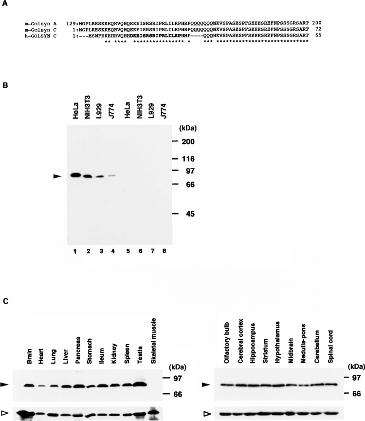
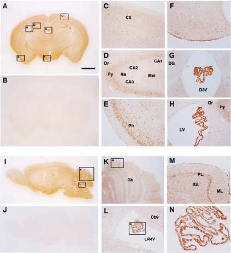
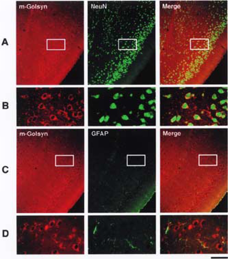
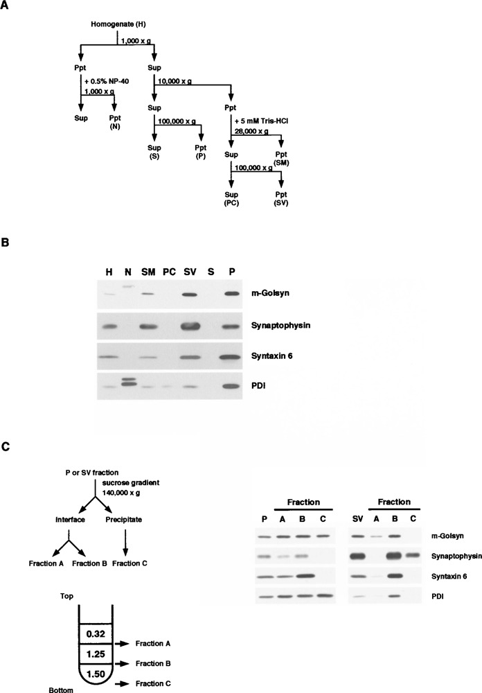
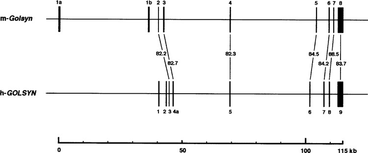
The mouse ortholog of the human GOLSYN gene, termed the m-Golsyn gene, was isolated and mapped to the region on mouse chromosome 15B3.2 syntenic with human chromosome 8q23. Three mRNA species (type la, 1b, and type 2) were produced by use of alternative transcription initiation points and alternative splicing events. The type 1 mRNAs were expressed only in the brain, whereas the type 2 was detected in various tissues. m-Golsyn protein was expressed in various tissues including the brain. Immunohistochemical study of m-Golsyn protein showed its prominent expression in the neuronal cells in various regions of the brain and strong expression in the choroid plexus ependymal cells lining the ventricles. m-Golsyn protein was found to be homologous to syntaphilin, a regulator of synaptic vesicle exocytosis. These results indicate that the m-Golsyn protein may play an important role in intracellular protein transport in neuronal cells of the brain.


Figures

Figure 1
Figure 1
Genomic, mRNA, and protein structures of the m-Golsyn gene. (A) Exon–intron structure of the m-Golsyn gene (mouse ortholog of the human-GOLSYN gene). The m-Golsyn gene consists of 8 exons, and is situated within a 115-kb region of the RP24-288B1 BAC clone that is mapped to the mouse chromosome 15B3.2 region. The arrow pointing to the right indicates the position and the direction of m-Golsyn gene. Segments of EST (GenBank Accession Nos. BB647074, BM951171, BM947901, and BB023907), which were used for the determination of the genomic structure of the m-Golsyn gene, are indicated. Boxes indicate the position of exons. (B) Structure of the three types of m-Golsyn mRNA. Thick horizontal lines under each transcript indicate the position of the open reading frame. (C) Deduced amino acid sequences of the three m-Golsyn proteins. Their functional domains were predicted from the results of the functional search of a database: a potential coiled-coil domain is boxed, and a putative transmembrane domain is shaded.
Figure 2
Figure 2
Expression analysis of m-Golsyn transcripts in mouse tissues. cDNA fragments of m-Golsyn type 1 and type 2 were amplified by using the primer sets described under Materials and Methods. (A) The positions of the two sets of primers are indicated by the arrows. (B) The cDNA fragments of m-Golsyn type 1 and type 2 were amplified by using first-strand cDNAs derived from nine different mouse tissues. Lane 1 in (B) is a negative control lacking template cDNA. PCR was performed with cDNA derived from 0.15 μg of RNA for the amplification of m-Golsyn cDNAs and mouse actin cDNA.
Figure 3
Figure 3
Expression of m-Golsyn protein in mouse cell lines and tissues. (A) Parts of the peptide sequences of m-Golsyn and h-GOLSYN are indicated. The peptide sequence in bold type was used for the preparation of the anti-h-GOLSYN antibody. (B) Extracts (20 μg/lane) of HeLa, NIH3T3, L929, and J774 cells were separated by SDS-PAGE on a 7.5% slab gel and immunoblotted with anti-GOLSYN antibody in the absence (lanes 1–4) or presence (lanes 5–8) of an excess amount of the peptide used for the preparation of anti-GOLSYN antibody. (C) Lysates (50 μg/lane) prepared from various mouse tissues were separated on a 7.5% SDS-PAGE and analyzed by immunoblotting with anti-GOLSYN antibody as described in Materials and Methods. Left panel: various tissues including brain; right panel: various regions of the central nervous system. Solid and open arrowheads indicate the positions of m-Golsyn protein and β-tubulin, respectively.
Figure 4
Figure 4
Immunohistochemical analysis of m-Golsyn protein in mouse brain. Coronal (A–H) and sagittal (I–N) sections of the mouse brain were stained with anti-GOLSYN antibody in the presence (B and J) or absence (A to N except B and J) of an excess amount of the peptide used for the preparation of anti-GOLSYN antibody. Regions indicated by boxes C to H in (A) are magnified and shown in (C) to (H), respectively. Boxes K and L in (I) are magnified and shown in (K) and (L), respectively. CX, cerebral cortex; Or, oriens layer of the hippocampus; Py, pyramidal cell layer of the hippocampus; CA1, CA2, and CA3, CA1, CA2, and CA3 areas of the hippocampus; Ra, stratum radiatum of the hippocampus; Mol, molecular cell layer of the hippocampus; Pir, pirform cortex; DG, dendate gyrus; D3V, third ventricle; LV, lateral ventricle; Cb, cerebellum; LR4V, fourth ventricle; PL, Purkinje cell layer of the cerebellum; ML, molecular layer of cerebellum; IGL, inner granular layer of cerebellum. Scale bar: 1 mm.
Figure 5
Figure 5
Specific expression of m-Golsyn protein in neuronal cells of mouse brain. Localization of m-Golsyn, NeuN, and GFAP in mouse piriform cortex was analyzed as described in Materials and Methods. Enlarged photographs of the boxed regions in (A) and (C) are shown in (B) and (D), respectively. m-Golsyn and NeuN or GFAP are displayed in red and green, respectively. Scale bar: 135 μm (A, C) and 30 μm (B, D).
Figure 6
Figure 6
Subcellular distribution of m-Golsyn protein in mouse cerebral cortex. (A) Procedure for subcellular fractionation of mouse cerebral cortex. Homogenates were separated into fractions enriched in nuclei (N), 100,000 × g supernatant (S), 100,000 × g precipitate (P), synaptic plasma membrane (SM), presynaptic cytosol (PC), and synaptic vesicle (SV) by differential centrifugation. (B) Fractions were prepared as shown in (A), and an aliquot (20 μg protein) of each of these fractions was resolved by 7.5% SDS-PAGE and subjected to immunoblot analysis with antibodies against GOLSYN, synaptophysin, syntaxin 6, or PDI. Synaptophysin, syntaxin 6, and PDI were used as markers for synaptic vesicle, Golgi apparatus, and endoplasmic reticulum, respectively. Solid and gray arrowheads indicate the position of m-Golsyn and PDI, respectively. (C) P and SV fractions, prepared as shown in (A), were fractionated by ultracentrifugation of a sucrose gradient and subjected to immunoblot analysis.
Figure 7
Figure 7
Schematic representation of exon–intron structure of m-Golsyn and h-GOLSYN gene. Boxes indicate the position of exons. Horizontal lines interconnecting exons represent introns. The numbers indicate the similarity between m-Golsyn and h-GOLSYN gene at the nucleotide sequence level.

Similar articles

Cited by

References

    1. Altschul S. F.; Gish W.; Miller W.; Myers E. W.; Lipman D. J. Basic local alignment search tool. J. Mol. Biol. 215:403–410; 1990. - PubMed
    1. Chakrabortty S.; Kitada M.; Matsumoto N.; Taketomi M.; Kimura K.; Ide C. Choroid plexus ependymal cells enhance neurite outgrowth from dorsal root ganglion neurons in vitro. J. Neurocytol. 29:707–717; 2000. - PubMed
    1. Davson H. A comparative study of the aqueous humour and cerebrospinal fluid in the rabbit. J. Physiol. 129:111–133; 1955. - PMC - PubMed
    1. Debus E.; Weber K.; Osborn M. Monoclonal antibodies specific for glial fibrillary acidic (GFA) protein and for each of the neurofilament triplet polypeptides. Differentiation 25:193–203; 1983. - PubMed
    1. Dynan W. S.; Tjian R. Control of eukaryotic messenger RNA synthesis by sequence-specific DNA-binding proteins. Nature 316:774–778; 1985. - PubMed

Publication types

MeSH terms

LinkOut - more resources