Comparative Study
. 2006 Spring;11(1):13-22.
doi: 10.1379/csc-138r.1.

Toxoplasma gondii-derived heat shock protein 70 stimulates maturation of murine bone marrow-derived dendritic cells via Toll-like receptor 4

Affiliations
Comparative Study

Toxoplasma gondii-derived heat shock protein 70 stimulates maturation of murine bone marrow-derived dendritic cells via Toll-like receptor 4

Fumie Aosai et al. Cell Stress Chaperones. 2006 Spring.

Abstract

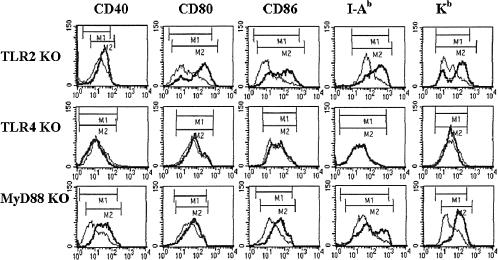
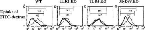
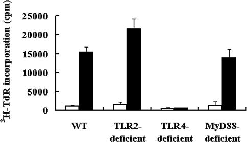
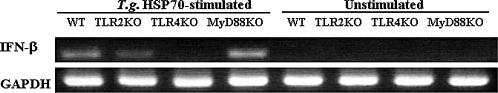
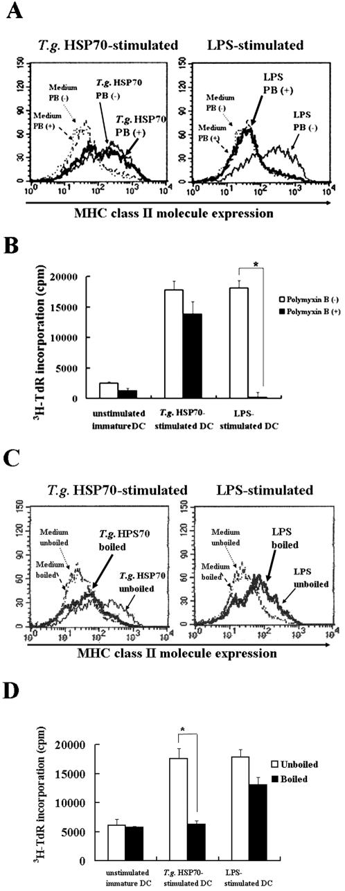
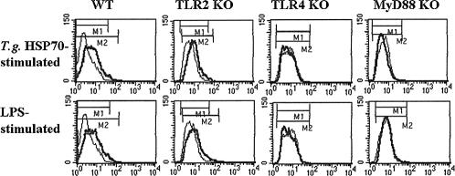
Toxoplasma gondii-derived heat shock protein 70 (T.g.HSP70) induced maturation of bone marrow-derived dendritic cells (DCs) of wild-type (WT) C57BL/6 mice as evidenced by an increase in surface expression of MHC class I and II molecules and costimulatory molecules such as CD40, CD80, and CD86. Functionally, decreased phagocytic ability and increased alloreactive T cell stimulatory ability were observed in T.g.HSP70-stimulated DCs. These phenotypic and functional changes of T.g.HSP70-stimulated DCs were demonstrated in Toll-like receptor (TLR) 2- and myeloid differentiation factor 88 (MyD88)-deficient but not TLR4-deficient C57BL/6 mice. DCs from WT and TLR2-deficient but not TLR4-deficient mice produced IL-12 after T.g.HSP70 stimulation. T.g.HSP70-stimulated DCs from WT, TLR2-deficient, and MyD88-deficient, but not TLR4-deficient mice expressed IFN-beta mRNA. Thus, T.g.HSP70 stimulates murine DC maturation via TLR4 through the MyD88-independent signal transduction cascade.


Figures

Fig 1.
Fig 1.
(A) Flow cytometric analyses of CD40, CD80, CD86, and MHC expression on T.g.HSP70-stimulated BMDCs. Immature BMDCs from WT mice were cultured with or without 1 μg/ml T.g.HSP70 or 100 ng/ml LPS for 48 hours and stained with PE-conjugated anti-CD40, -CD80, or -CD86 mAb, or FITC-conjugated anti-I-Ab or -Kb mAb. Surface expressions of the costimulatory molecules (CD40, CD80, and CD86) and MHC class I and II molecules of BMDCs stimulated with (bold lines) or without (thin lines) T.g.HSP70 or LPS were comparatively analyzed by FACScan. Representative data from three independent experiments are shown. (B) Dose-response analysis. Immature BMDCs from WT mice were cultured with the indicated amounts of T.g.HSP70 (black bars) or LPS (striped bars), or medium alone (open bar) for 48 hours. Surface expressions of CD86 were shown as mean fluorescence intensities. Differences of fluorescence intensities between T.g.HSP70- or LPS-stimulated and unstimulated BMDCs were determined by Kolmogorov-Smirnov (K-S) two-sample test. *, P < 0.001
Fig 2.
Fig 2.
Involvement of TLR in T.g.HSP70-stimulated BMDC maturation. Immature BMDCs from TLR2-, TLR4-, and MyD88-deficient B6 mice were cultured with or without T.g.HSP70 for 48 hours, and surface expressions of CD40, CD80, CD86, and MHC class I and II molecules of BMDCs cultured with (bold lines) or without (thin lines) T.g.HSP70 were comparatively analyzed by FACScan. Representative data from three independent experiments are shown
Fig 3.
Fig 3.
Uptake of FITC-dextran by T.g.HSP70-stimulated BMDCs. Immature BMDCs from WT, TLR2-deficient, TLR4-deficient, and MyD88-deficient B6 mice were cultured with (bold lines) or without (thin lines) T.g.HSP70 for 48 hours and were incubated in a 96-well microtiter plate with FITC-dextran at a concentration of 1 mg/ml for 1 hour at 37°C. Uptake of FITC-dextran by BMDCs was analyzed by FACScan. Representative data from three independent experiments are shown
Fig 4.
Fig 4.
Alloreactive T cell stimulatory activity by T.g.HSP70-stimulated BMDCs. Immature BMDCs from WT, TLR2-deficient, TLR4-deficient, and MyD88-deficient B6 mice were cultured with (filled bars) or without (open bars) T.g.HSP70 for 48 hours and irradiated with 20 Gy. 1 × 10⁴ BMDCs from each mouse strain were cocultured as stimulator cells with 1 × 10⁵ responder T cells from spleens of BALB/c mice. Proliferation of responder cells is expressed as counts per minute (cpm) of [3H]thymidine incorporation. Data are representative of three independent experiments
Fig 5.
Fig 5.
IFN-β mRNA expression of T.g.HSP70-stimulated BMDCs. Immature BMDCs from WT (lanes 1 and 5), TLR2-deficient (lanes 2 and 6), TLR4-deficient (lanes 3 and 7), and MyD88-deficient (lanes 4 and 8) B6 mice were cultured with T.g.HSP70 for 48 hours, and expressions of IFN-β mRNA in BMDCs stimulated with (lanes 1–4) or without (lanes 5–8) T.g.HSP70 were investigated by RT-PCR. The sequences of forward and reverse IFN-β primers used were 5′-TCCAAGAAAGGACGAACATTCG-3′ and 5′-TGAGGACATCTC CCACGTCAA-3′, respectively. GAPDH was used for internal control. Data are representative of three independent experiments
Fig 6.
Fig 6.
Specificity of T.g.HSP70 to stimulate DC maturation. Effects of Polymyxin B on up-regulation of MHC class II molecule expression (A) and on alloreactive T cell stimulatory ability (B) of T.g.HSP70- or LPS-stimulated BMDCs were examined by adding 10 μg/ml Polymyxin B to the culture of immature BMDCs of WT mice with or without T.g.HSP70 or LPS. Effects of boiled (100°C, 1 hour) T.g.HSP70 or LPS on immature BMDCs to induce surface MHC class II molecule expression (C) or alloreactive T cell stimulatory ability (D) were also examined. In FACScan analysis (A and C), MHC class II molecule expressions of BMDCs are shown as follows: medium without Polymyxin B or boiling (thin dotted lines), medium with Polymyxin B or boiling (bold dotted lines), T.g.HSP70- or LPS-stimulated without Polymyxin B or boiling (thin solid lines) and T.g.HSP70- or LPS-stimulated with Polymyxin B or boiling (bold solid lines). In alloreactive T cell stimulation (B and D), cultures with (filled bars) or without (open bars) treatment by Polymyxin B or boiling were compared. Differences of mean values between untreated and treated groups were determined by unpaired Student's t-test. *, P < 0.01; cpm, counts per minute. Data are representative of three separate experiments
Fig 7.
Fig 7.
IL-12 production by T.g.HSP70-stimulated BMDCs. Immature BMDCs from WT, TLR2-deficient, TLR4-deficient, and MyD88-deficient B6 mice were cultured with (bold lines) or without (thin lines) T.g.HSP70 or LPS for 48 hours and stained with PE-conjugated anti-IL-12 mAb by Cytofix/Cytoperm kit (PharMingen). After washing, the intracellular expression of IL-12 of BMDCs was analyzed by FACScan. Representative data from three independent experiments are shown

Similar articles

Cited by

References

    1. Adachi O, Kawai T, and Takeda K. et al. 1998 Targeted disruption of the MyD88 gene results in loss of IL-1- and IL-18-mediated function. Immunity. 9:143–150. - PubMed
    1. Ahmed AK, Mun HS, Aosai F, Piao LX, Fang H, Norose K, Yano A. Roles of Toxoplasma gondii-derived heat shock protein 70 in host defense against T. gondii infection. Microbiol Immunol. 2004;48:911–915.0385-5600(2004)048[0911:ROTGHS]2.0.CO;2 - PubMed
    1. Akira S, Takeda K. Toll-like receptor signaling. Nat Rev Immunol. 2004;4:499–511.1474-1733(2004)004[0499:TRS]2.0.CO;2 - PubMed
    1. Aliberti J, Jankovic D, Sher A. Turning it on and off: regulation of dendritic cell function in Toxoplasma gondii infection. Immunol Rev. 2004;201:26–34.0105-2896(2004)201[0026:TIOAOR]2.0.CO;2 - PubMed
    1. Aliberti J, Reis e Sousa C, Schito M, Hieny S, Wells T, Huffnagle GB, Sher A. CCR5 provides a signal for microbial induced production of IL-12 by CD8α+ dendritic cells. Nat Immunol. 2000;1:83–87.1529-2908(2000)001[0083:CPASFM]2.0.CO;2 - PubMed

Publication types

Substances

LinkOut - more resources