

Binge alcohol treatment increases vertebral bone loss following ovariectomy: compensation by intermittent parathyroid hormone
- PMID: 16573585
- PMCID: PMC3065175
- DOI: 10.1111/j.1530-0277.2006.00078.x
Binge alcohol treatment increases vertebral bone loss following ovariectomy: compensation by intermittent parathyroid hormone
Abstract
Background: Postmenopausal estrogen deficiency and alcohol abuse are known risk factors for osteoporosis. Previous studies of the combined effect of alcohol and ovariectomy on bone loss using chronic alcohol-feeding models have not demonstrated additional alcohol-induced bone loss in ovariectomized (OVX) animals. Binge alcohol treatment causes rapid bone loss in male rats. We hypothesized that binge alcohol would cause additional bone loss in OVX rats.
Methods: Ninety-six adult (400 g) female Sprague-Dawley rats (48 sham-operated and 48 OVX, pair fed) were randomly divided into 4 treatment groups: (a) saline-treated, (b) binge alcohol-treated (3 g/kg alcohol as a 20% weight to volume alcohol/saline solution, intraperitoneal (IP), 3 times per week), (c) parathyroid hormone (PTH)-treated (80 microg/kg, SC, 5 d/wk), and (d) binge alcohol plus PTH. Rats were treated for either 2 or 4 weeks. Following treatment periods, blood was collected for alcohol concentration (BAC) measurements; lumbar vertebrae were removed for bone mineral density (BMD) levels, trabecular microarchitecture assessment, and vertebral compressive strength analysis.
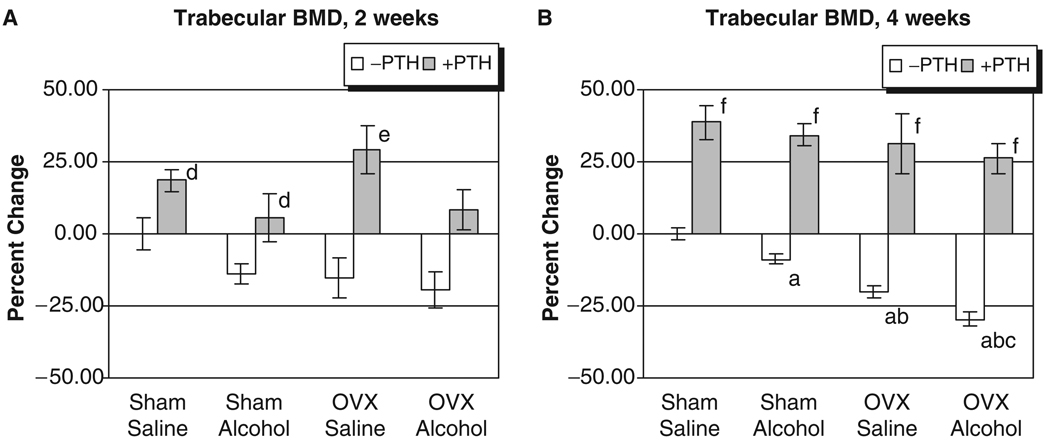
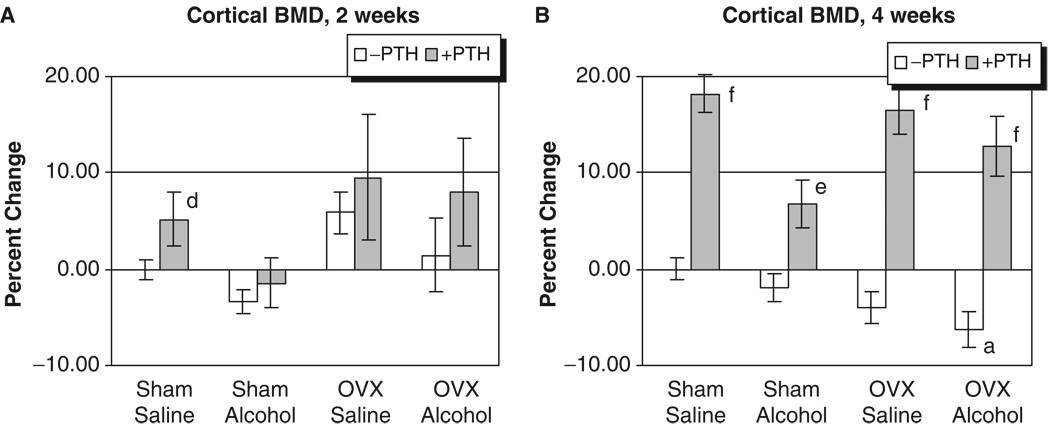
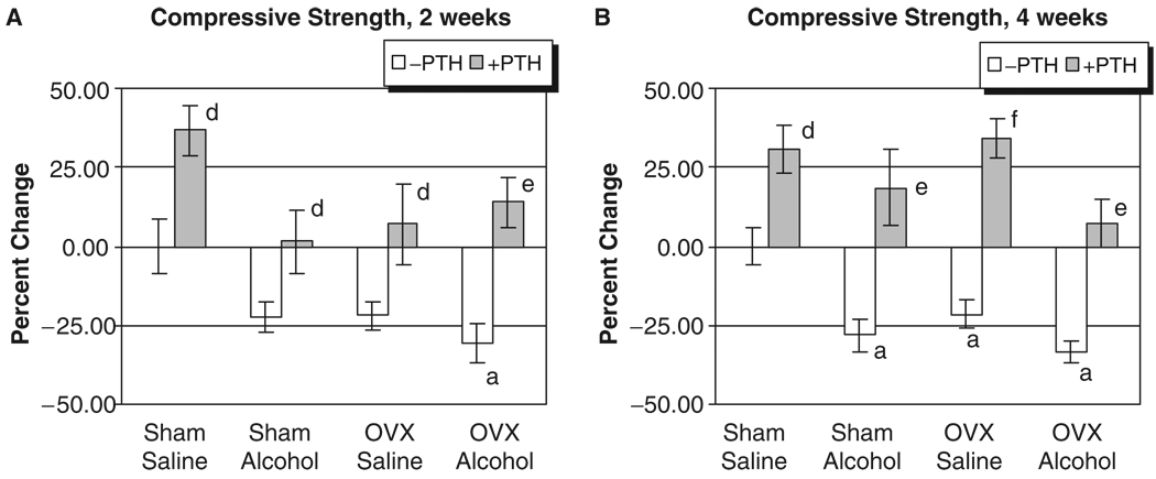
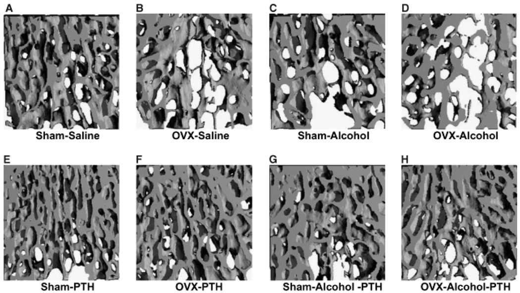
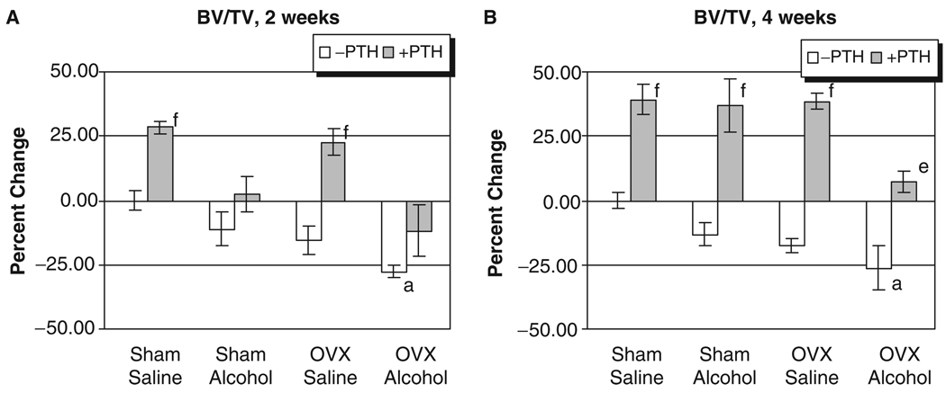
Results: Peak binge BACs averaged 300 mg/dL. Alcohol and OVX decreased cancellous BMD: alcohol and OVX treatment in combination caused additional cancellous BMD loss and significant cortical BMD reductions. Compressive strength was also decreased by OVX and alcohol. Combination treatment resulted in further declines in bone strength. Micro-CT analysis revealed a significant effect of combined OVX and alcohol treatment resulting in decreased trabecular bone volume/total volume (BV/TV). Intermittent PTH administration compensated for losses of BMD, compressive strength, and restored BV/TV deficits caused by OVX, alcohol, or their combination.
Conclusions: Bone loss following OVX can be significantly increased by concurrent binge alcohol treatment. The effects of alcohol and OVX are compensated by concurrent intermittent treatment with PTH. These results suggest that postmenopausal women who abuse alcohol may place their skeleton at additional risk for osteoporotic fracture.
Figures
References
-
- Angus RM, Sambrook PN, Pocock NA. Dietary intake and bone mineral density. Osteoporosis Int. 1988;1:265–277. - PubMed
-
- Bilke DD, Genant HK, Cann C, Recker RR, Halloran BP, Strewler GJ. Bone disease in alcohol abuse. Ann Intern Med. 1985;103:42–48. - PubMed
-
- Blumenthal SJ. Women and substance abuse: a new national focus. In: Wetherington CL, Roman AB, editors. Drug Addiction Research and the Health of Women. Bethesda, MD: USDHHS, NIH/NIDA; 1998. pp. 13–32.
-
- Cavolina JM, Evans GL, Harris SA, Zhang M, Westerlind KC, Turner RT. The effects of orbital spaceflight on bone histomorphometry and messenger ribonucleic acid levels for bone matrix proteins and skeletal signaling peptides in ovariectomized growing rats. Endocrinology. 1997;8:1567–1576. - PubMed
Publication types
MeSH terms
Substances
Grants and funding
LinkOut - more resources
Full Text Sources
Medical
