. 2006 Mar 30:4:15.
doi: 10.1186/1479-5876-4-15.

Innate immune functions of microglia isolated from human glioma patients

Affiliations

Innate immune functions of microglia isolated from human glioma patients

S Farzana Hussain et al. J Transl Med. .

Abstract

Background: Innate immunity is considered the first line of host defense and microglia presumably play a critical role in mediating potent innate immune responses to traumatic and infectious challenges in the human brain. Fundamental impairments of the adaptive immune system in glioma patients have been investigated; however, it is unknown whether microglia are capable of innate immunity and subsequent adaptive anti-tumor immune responses within the immunosuppressive tumor micro-environment of human glioma patients. We therefore undertook a novel characterization of the innate immune phenotype and function of freshly isolated human glioma-infiltrating microglia (GIM).

Methods: GIM were isolated by sequential Percoll purification from patient tumors immediately after surgical resection. Flow cytometry, phagocytosis and tumor cytotoxicity assays were used to analyze the phenotype and function of these cells.

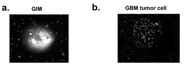
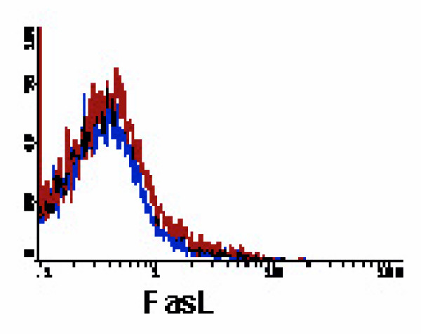
Results: GIM expressed significant levels of Toll-like receptors (TLRs), however they do not secrete any of the cytokines (IL-1beta, IL-6, TNF-alpha) critical in developing effective innate immune responses. Similar to innate macrophage functions, GIM can mediate phagocytosis and non-MHC restricted cytotoxicity. However, they were statistically less able to mediate tumor cytotoxicity compared to microglia isolated from normal brain. In addition, the expression of Fas ligand (FasL) was low to absent, indicating that apoptosis of the incoming lymphocyte population may not be a predominant mode of immunosuppression by microglia.

Conclusion: We show for the first time that despite the immunosuppressive environment of human gliomas, GIM are capable of innate immune responses such as phagocytosis, cytotoxicity and TLR expression but yet are not competent in secreting key cytokines. Further understanding of these innate immune functions could play a critical role in understanding and developing effective immunotherapies to malignant human gliomas.


Figures

Figure 1
Figure 1
Isolation of GIM and expression of surface markers demonstrating the purity of the GIM population (CD11b vs. CD45) at each isolated fraction. Each interphase from the second percoll gradient was analyzed for expression of the surface markers CD11b and CD45. The gated cells in the upper right quadrant indicate the percentage of CD45+ gated cells that are also positive for CD11b and represent GIM. An autofluorescent population was identified in both normal and GBM tissue samples during flow cytometry analysis and is also consistently present in the respective isotype controls. This population was excluded when calculating the percentages of positive fluorescing cells.
Figure 2
Figure 2
GIM express TLRs. GIM purity was determined as in Fig. 1 and the cells were double-stained for CD45 and TLRs. All CD45+ expressing cells were gated and observed for TLR expression. In each histogram, the data plot on the left indicates the isotype control, and the second plot represents CD45+ gated cells that expressed the respective TLR. These data are from one tumor specimen but are representative of at least five tumor tissue samples from human glioma patients.
Figure 3
Figure 3
GIM do not secret innate cytokines. GIM purity was determined as in Fig. 1 and the cells were stained for intracellular cytokines IFN-α, IL-6, TNF-α. In each histogram, the data plot on the left indicates the isotype control, and the second plot represents respective cytokine producing cells. These data are from one tumor specimen but are representative of seven tumor tissue samples from human glioma patients and A375 control cells.
Figure 4
Figure 4
GIM can mediate phagocytosis. Cells were incubated with opsonized fluorescent beads and analyzed using fluorescence microscopy at 20× magnification. A, GIM containing phagocytosed beads. B, Control tumor cell that has fluorescent beads sticking to the surface but not internalized. Cells were analyzed through a z-stack to determine whether the beads were internalized or merely on the surface.
Figure 5
Figure 5
GIM can mediate non-MHC-restricted tumor cytotoxicity. GIM, peripheral blood mononuclear cells, and normal (5 × 10⁶, 1 × 10⁶, or 5 × 10⁵) microglia were each incubated as an effector population with 1 × 10⁵ carboxy-fluorescein diacetate succinimidyl ester-labeled U-87 target cells, for an effector-to-target ratio of 50:1, 10:1, or 5:1, respectively. Propidium iodide was added after 24 hours incubation, and cells were analyzed by flow cytometry. Cells were first gated only on the carboxy-fluorescein diacetate succinimidyl ester+ target population (excluding all other cells in the assay), and the propidium iodide expression on these cells was determined. The percentages of cytolytic activity of the effector cells were calculated (Y axis)(dead targets in upper right quadrant/[dead targets in upper right quadrant + live targets in lower right quadrant]) and plotted against each respective effector-to-target ratio (X axis). These data are from one tumor specimen and are representative of experiments from cells isolated from 3 different glioma patients. Bars at each data point represent 95% confidence intervals for each proportion. After 24 hours, GIM were functional in their tumor cytotoxic activity and comparable to PBMCs, however cytotoxic activity of GIM was significantly (*p < 0.0001) lower than that of normal microglia.
Figure 6
Figure 6
GIM cannot initiate Fas-mediated apoptosis. The histogram is depicted similar to that in Fig. 2. GIM were gated on CD45+ expression and were analyzed for expression of FasL. These data are from one tumor specimen and are representative of at least 11 tumor tissue samples from human glioma patients.

Similar articles

Cited by

References

    1. Davis FG, Kupelian V, Freels S, McCarthy B, Surawicz T. Prevalence estimates for primary brain tumors in the United States by behavior and major histology groups. Neuro-oncol. 2001;3:152–158. - PMC - PubMed
    1. Ridley A. Immunological aspects of gliomas. Proc R Soc Med. 1977;70:867–869. - PMC - PubMed
    1. Holladay FP, Choudhuri R, Heitz T, Wood GW. Generation of cytotoxic immune responses during the progression of a rat glioma. J Neurosurg. 1994;80:90–96. - PubMed
    1. Fathallah-Shaykh HM, Gao W, Cho M, Herrera MA. Priming in the brain, an immunologically privileged organ, elicits anti-tumor immunity. Int J Cancer. 1998;75:266–276. - PubMed
    1. Merchant RE, Grant AJ, Merchant LH, Young HF. Adoptive immunotherapy for recurrent glioblastoma multiforme using lymphokine activated killer cells and recombinant interleukin-2. Cancer. 1988;62:665–671. - PubMed