

Postfatigue potentiation of the paralyzed soleus muscle: evidence for adaptation with long-term electrical stimulation training
- PMID: 16575026
- PMCID: PMC3270308
- DOI: 10.1152/japplphysiol.00099.2006
Postfatigue potentiation of the paralyzed soleus muscle: evidence for adaptation with long-term electrical stimulation training
Abstract
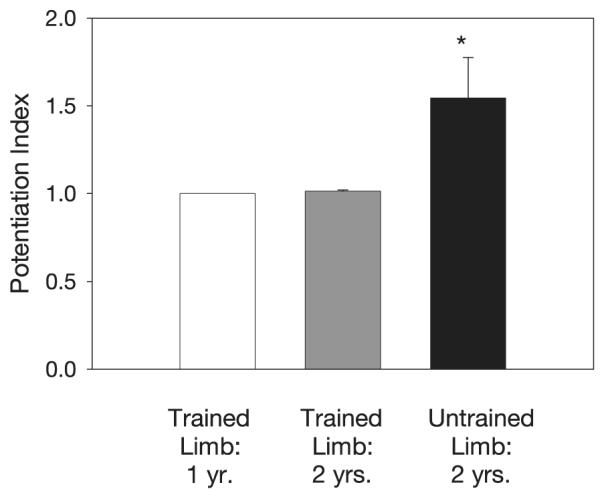
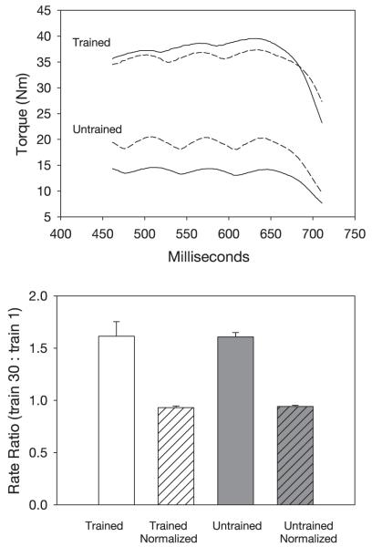
Understanding the torque output behavior of paralyzed muscle has important implications for the use of functional neuromuscular electrical stimulation systems. Postfatigue potentiation is an augmentation of peak muscle torque during repetitive activation after a fatigue protocol. The purposes of this study were 1) to quantify postfatigue potentiation in the acutely and chronically paralyzed soleus and 2) to determine the effect of long-term soleus electrical stimulation training on the potentiation characteristics of recently paralyzed soleus muscle. Five subjects with chronic paralysis (>2 yr) demonstrated significant postfatigue potentiation during a repetitive soleus activation protocol that induced low-frequency fatigue. Ten subjects with acute paralysis (<6 mo) demonstrated no torque potentiation in response to repetitive stimulation. Seven of these acute subjects completed 2 yr of home-based isometric soleus electrical stimulation training of one limb (compliance = 83%; 8,300 contractions/wk). With the early implementation of electrically stimulated training, potentiation characteristics of trained soleus muscles were preserved as in the acute postinjury state. In contrast, untrained limbs showed marked postfatigue potentiation at 2 yr after spinal cord injury (SCI). A single acute SCI subject who was followed longitudinally developed potentiation characteristics very similar to the untrained limbs of the training subjects. The results of the present investigation support that postfatigue potentiation is a characteristic of fast-fatigable muscle and can be prevented by timely neuromuscular electrical stimulation training. Potentiation is an important consideration in the design of functional electrical stimulation control systems for people with SCI.
Figures


Similar articles
-
Musculoskeletal adaptations in chronic spinal cord injury: effects of long-term soleus electrical stimulation training.Neurorehabil Neural Repair. 2007 Mar-Apr;21(2):169-79. doi: 10.1177/1545968306293447. Neurorehabil Neural Repair. 2007. PMID: 17312092 Free PMC article.
-
Musculoskeletal plasticity after acute spinal cord injury: effects of long-term neuromuscular electrical stimulation training.J Neurophysiol. 2006 Apr;95(4):2380-90. doi: 10.1152/jn.01181.2005. Epub 2006 Jan 11. J Neurophysiol. 2006. PMID: 16407424 Free PMC article. Clinical Trial.
-
Doublet electrical stimulation enhances torque production in people with spinal cord injury.Neurorehabil Neural Repair. 2011 Jun;25(5):423-32. doi: 10.1177/1545968310390224. Epub 2011 Feb 8. Neurorehabil Neural Repair. 2011. PMID: 21304018 Free PMC article.
-
Effect of muscle length on maximum evoked torque, discomfort, contraction fatigue, and strength adaptations during electrical stimulation in adult populations: A systematic review.PLoS One. 2024 Jun 10;19(6):e0304205. doi: 10.1371/journal.pone.0304205. eCollection 2024. PLoS One. 2024. PMID: 38857245 Free PMC article.
-
Muscle and bone plasticity after spinal cord injury: review of adaptations to disuse and to electrical muscle stimulation.J Rehabil Res Dev. 2008;45(2):283-96. doi: 10.1682/jrrd.2007.02.0031. J Rehabil Res Dev. 2008. PMID: 18566946 Free PMC article. Review.
Cited by
-
Low force contractions induce fatigue consistent with muscle mRNA expression in people with spinal cord injury.Physiol Rep. 2014 Feb 25;2(2):e00248. doi: 10.1002/phy2.248. eCollection 2014 Feb 1. Physiol Rep. 2014. PMID: 24744911 Free PMC article.
-
Doublet stimulation protocol to minimize musculoskeletal stress during paralyzed quadriceps muscle testing.J Appl Physiol (1985). 2008 Jun;104(6):1574-82. doi: 10.1152/japplphysiol.00892.2007. Epub 2008 Apr 24. J Appl Physiol (1985). 2008. PMID: 18436697 Free PMC article.
-
Repeated maximal volitional effort contractions in human spinal cord injury: initial torque increases and reduced fatigue.Neurorehabil Neural Repair. 2009 Nov;23(9):928-38. doi: 10.1177/1545968309336147. Epub 2009 May 28. Neurorehabil Neural Repair. 2009. PMID: 19478056 Free PMC article.
-
RNA-seq data of soleus muscle tissue after spinal cord injury under conditions of inactivity and applied exercise.Data Brief. 2019 Dec 31;28:105056. doi: 10.1016/j.dib.2019.105056. eCollection 2020 Feb. Data Brief. 2019. PMID: 32226812 Free PMC article.
-
Dose estimation and surveillance of mechanical loading interventions for bone loss after spinal cord injury.Phys Ther. 2008 Mar;88(3):387-96. doi: 10.2522/ptj.20070224. Epub 2008 Jan 17. Phys Ther. 2008. PMID: 18202080 Free PMC article.
References
-
- American Spinal Injury Association . International Standards for Neurological Classification of SCI. American Spinal Injury Association; Atlanta, GA: 2002.
-
- Baldwin KM, Roy RR, Sacks RD, Blanco C, Edgerton VR. Relative independence of metabolic enzymes and neuromuscular activity. J Appl Physiol. 1984;56:1602–1607. - PubMed
-
- Bozzo C, Spolaore B, Toniolo L, Stevens L, Bastide B, Cieniewski-Bernard C, Fontana A, Mounier Y, Reggiani C. Nerve influence on myosin light chain phosphorylation in slow and fast skeletal muscles. FEBS J. 2005;272:5771–5785. - PubMed
-
- Bozzo C, Stevens L, Toniolo L, Mounier Y, Reggiani C. Increased phosphorylation of myosin light chain associated with slow-to-fast transition in rat soleus. Am J Physiol Cell Physiol. 2003;285:C575–C583. - PubMed
Publication types
MeSH terms
Grants and funding
LinkOut - more resources
Full Text Sources
Medical