. 2006 Apr 1;78(7):2190-8.
doi: 10.1021/ac051709x.

Enhanced mixture analysis of poly(ethylene glycol) using high-field asymmetric waveform ion mobility spectrometry combined with fourier transform ion cyclotron resonance mass spectrometry

Affiliations

Enhanced mixture analysis of poly(ethylene glycol) using high-field asymmetric waveform ion mobility spectrometry combined with fourier transform ion cyclotron resonance mass spectrometry

Errol W Robinson et al. Anal Chem. .

Abstract

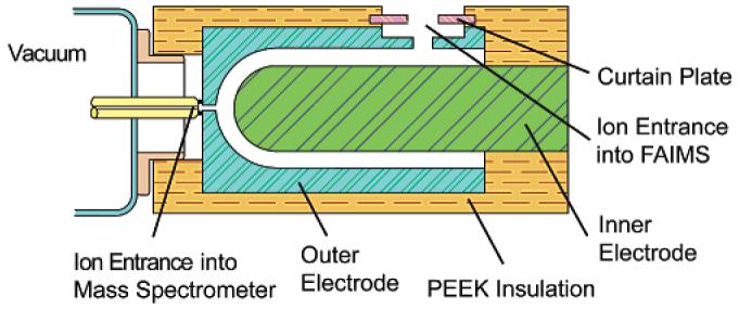
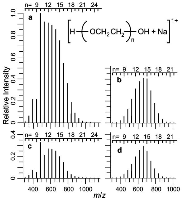
The combination of high-field asymmetric waveform ion mobility spectrometry (FAIMS) with Fourier transform ion cyclotron resonance mass spectrometry (FTICR-MS) makes possible lower detection limits, increased sensitivity, and increased dynamic range in the analysis of poly(ethylene glycol) (PEG) samples of low molecular weight. The signal gain obtained using FAIMS depends on ion identity, with a range between 1.8x and 14x obtained for various molecular ions of PEG 600. A 1.7-fold reduction in noise is obtained using FAIMS due to the elimination of chemical noise. The improved detection performance is predominantly due to a reduction in adverse Coulomb effects as a result of ions being selectively introduced into the mass spectrometer. The high ion transmission obtained using FAIMS combined with the high sensitivity of FTICR-MS detection make possible separation of multiple gas-phase conformers of PEG molecular cations that have low abundance (less than 0.2% relative abundance) and that have not been detected previously. Mixed dications of PEG that have the same nominal mass but differ by the number polymer subunits (m/Delta m up to 25,000) can be separately introduced into the mass spectrometer using FAIMS. Interactions of the carrier gas with the metal ions that are attached to the PEG molecules appear to be the most significant factor in these FAIMS separations.


Figures

Figure 1
Figure 1
Schematic diagram of the FAIMS device attached to the ion entrance of the FTICR mass spectrometer.
Figure 2
Figure 2
Relative intensities of singly charged sodiated PEG 600 ions: (a) maximum intensity through the FAIMS device observed for each PEG n-mer, (b) maximum intensity observed for each n-mer without FAIMS from a set of 10 mass spectra, (c) average relative intensity observed for each n-mer with FAIMS summed over ∼10 spectra that contribute to a CV peak, and (d) average relative intensity observed for each n-mer without FAIMS (10-scan average). All relative intensities (a–d) are on the same scale. Note that the noise level on the 10-scan averages (c,d) are lower due to signal averaging. The enhanced ion abundances of PEG 7- and 10-mers in (c) are due to more selective transmission of these ions through the FAIMS device (see text).
Figure 3
Figure 3
Partial mass spectra of PEG 600 with FAIMS at compensation voltages of −7.4 (a) and −5.9 V (b). The ion intensities are plotted relative to the ion intensity in Figure 2a.
Figure 4
Figure 4
Mass spectra and CV scan for singly charged sodiated PEG with 22 polymer subunits: mass spectra without (a) and with (b) the FAIMS separation and (c) the CV scan for this ion. The ion intensities are plotted relative to the ion intensity in Figure 2a.
Figure 5
Figure 5
Compensation voltage scan of singly charged sodiated PEG with 14 polymer subunits: (a) a three-dimensional plot of CV versus partial mass spectra data around the region of the sodiated PEG 14-mer and (b) maximum relative conformer intensity versus CV. The major peak in transmission through the FAIMS device is labeled as region II. Minor peaks in ion transmission are labeled as regions I, III, and IV. The mass spectral data are not apodized so that the peak shape is Lorentzian. This results in broad peaks near the baseline (see green region in conformer II).
Figure 6
Figure 6
Major and minor peaks in ion transmission through the FAIMS device for singly charged sodiated PEG 600 with 5–27 polymer subunits. Smaller symbols correspond to low-abundance conformers.
Figure 7
Figure 7
Major peaks in ion transmission through the FAIMS device for singly charged PEG 600: (a) maximum compensation voltage of ion transmission versus number of polymer subunits and (b) maximum compensation voltage versus measured collisional cross sections. Lines marked with diamonds, triangles, and squares correspond to (PEG + Li)+, (PEG + Na)+, and (PEG + Cs)+, respectively. Collisional cross sections for lithiated, sodiated, and cesiated PEG ions are from previously reported values measured by Bowers and coworkers.-,,
Figure 8
Figure 8
Compensation voltage versus the number of incorporated polymer subunits for (PEG + 2Na)2+ (triangles), (PEG + Na + Cs)2+ (diamonds), and (PEG + 2Cs)2+ (squares), where the size of the symbol for each ion is proportional to the log of the absolute intensity for that ion.
Figure 9
Figure 9
Compensation voltage and mass spectra data for (PEG20 + 2H)2+, (PEG17 + Cs + H)2+, and (PEG19 + 2Na)2+: (a) a three-dimensional plot of CV versus m/z data (b) an overlay of three mass spectra, one for each ion obtained at three different CV values, and (c) CV scans for each ion.
Figure 10
Figure 10
Compensation voltage scan and mass spectra data for (PEG34 + 2H)2+ and (PEG33 + 2Na)2+ obtained from a PEG 1500 solution: (a) a three-dimensional plot of the CV versus m/z data (b) an overlay of two mass spectra, one for each ion obtained at two different CV values, and (c) an overlay of two CV scans, one for each ion.

References

    1. Shi SD-H, Hendrickson CL, Marshall AG. Proc. Natl. Acad. Sci. U.S.A. 1998;95:11532–11537. - PMC - PubMed
    1. He F, Hendrickson CL, Marshall AG. Anal. Chem. 2001;73:647–650. - PubMed
    1. Bossio RE, Marshall AG. Anal. Chem. 2002;74:1674–1679. - PubMed
    1. Mitchell DW, Smith RD. Phys. Rev. E. 1995;52:4366–4386. - PubMed
    1. Mitchell DW, Smith RD. J. Mass Spectrom. 1996;31:771–790.

Publication types