

Noninvasive Doppler tissue measurement of pulmonary artery compliance in children with pulmonary hypertension
- PMID: 16581479
- PMCID: PMC2003158
- DOI: 10.1016/j.echo.2005.11.012
Noninvasive Doppler tissue measurement of pulmonary artery compliance in children with pulmonary hypertension
Abstract
Background: We have shown previously that input impedance of the pulmonary vasculature provides a comprehensive characterization of right ventricular afterload by including compliance. However, impedance-based compliance assessment requires invasive measurements. Here, we develop and validate a noninvasive method to measure pulmonary artery (PA) compliance using ultrasound color M-mode (CMM) Doppler tissue imaging (DTI).
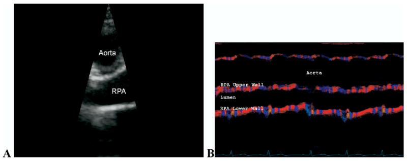
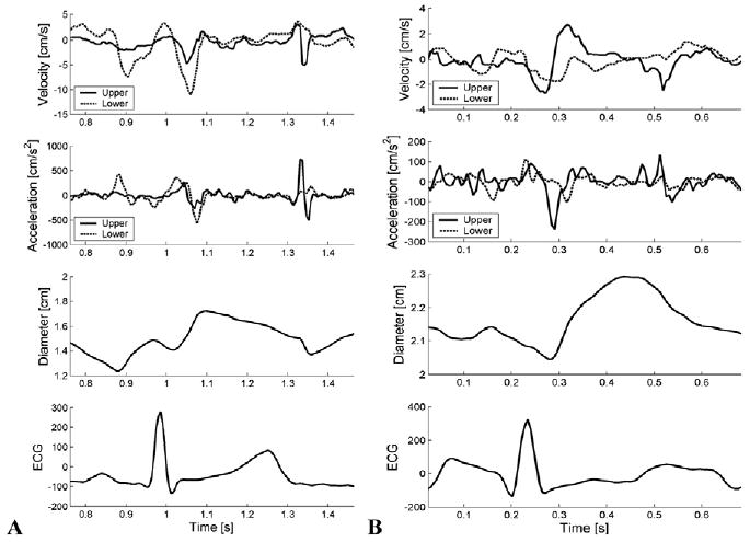
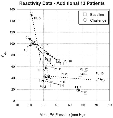
Methods: Dynamic compliance (C(dyn)) of the PA was obtained from CMM DTI and continuous wave Doppler measurement of the tricuspid regurgitant velocity. C(dyn) was calculated as: [(D(s) - D(d))/(D(d) x P(s))] x 10(4); where D(s) = systolic diameter, D(d) = diastolic diameter, and P(s) = systolic pressure. The method was validated both in vitro and in 13 patients in the catheterization laboratory, and then tested on 27 pediatric patients with pulmonary hypertension, with comparison with 10 age-matched control subjects. C(dyn) was also measured in an additional 13 patients undergoing reactivity studies.
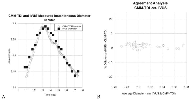
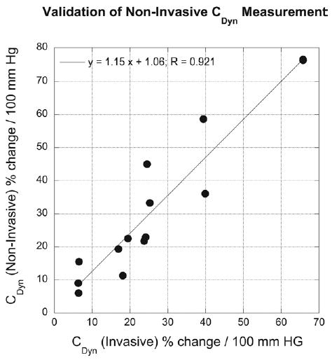
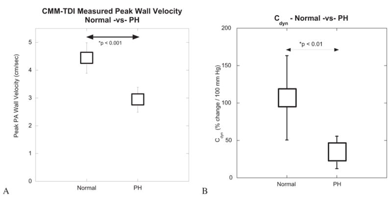
Results: Instantaneous diameter measured using CMM DTI agreed well with intravascular ultrasound measurements in the in vitro models. Clinically, C(dyn) calculated by CMM DTI agreed with C(dyn) calculated using invasive techniques (23.4 +/- 16.8 vs 29.1 +/- 20.6%/100 mm Hg; P = not significant). Patients with pulmonary hypertension had significantly lower peak wall velocity values and lower C(dyn) values than control subjects (P < .01). C(dyn) values followed an exponentially decaying relationship with PA pressure, indicating the nonlinear stress-strain behavior of these arteries. Reactivity in C(dyn) agreed with reactivity measured using impedance techniques.
Conclusion: The C(dyn) method provides a noninvasive means of assessing PA compliance and should be useful as an additional measure of vascular reactivity subsequent to pulmonary vascular resistance in patients with pulmonary hypertension.
Figures


References
-
- Weinberg C, Hertzberg J, Ivy DD, Kirby KS, Chen KC, Valdes-Cruz LM, et al. Extraction of pulmonary vascular compliance, PVR and RV work from single-pressure and Doppler flow measurements in children with pulmonary hypertension–a new method for evaluating reactivity: in vitro and clinical studies. Circulation. 2004;110:2609–17. - PubMed
-
- Weinberg C, Hertzberg J, Shandas R. Utility of intravascular ultrasound to measure local compliance of the pediatric pulmonary artery: in vitro studies. J Am Soc Echocardiogr. 2002;15:1507–14. - PubMed
-
- Press WH, Teukolsky SA, Vetterling WT, Flannery BP. Numerical recipes in C: the art of scientific computing. 2. New York: Cambridge University Press; 1992. p. 131.
-
- Berner M, Beghetti M, Spahr-Schopfer I, Oberhansli I, Friedli B. Inhaled nitric oxide to test the vasodilator capacity of the pulmonary vascular bed in children with long-standing pulmonary hypertension and congenital heart disease. Am J Cardiol. 1996;77:532–5. - PubMed
-
- Shandas R, Weinberg C, Ivy DD, Nicol E, DeGroff CG, Hertzberg J, et al. Development of a noninvasive ultrasound color M-mode means of estimating pulmonary vascular resistance in pediatric pulmonary hypertension: mathematical analysis, in vitro validation, and preliminary clinical studies. Circulation. 2001;104:908–14. - PubMed
Publication types
MeSH terms
Grants and funding
LinkOut - more resources
Full Text Sources
Medical
