Comparative Study
. 2006 Jun 15;573(Pt 3):857-67.
doi: 10.1113/jphysiol.2006.108274. Epub 2006 Mar 31.

Startle responses elicited by whiplash perturbations

Affiliations
Comparative Study

Startle responses elicited by whiplash perturbations

Jean-Sébastien Blouin et al. J Physiol. .

Abstract

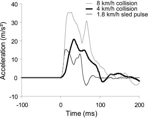
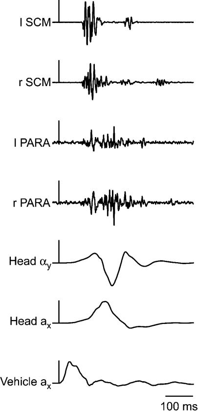
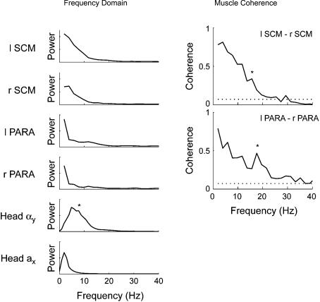
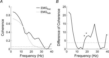
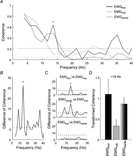
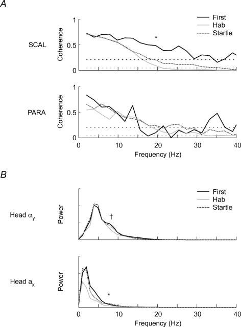
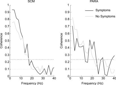
The human startle response produces muscle contractions throughout the body but the most brisk and synchronized contractions appear in the neck muscles. This response, which is greatest with the first exposure to a startling stimulus, could produce excessive and inappropriately directed muscle contractions that could explain the higher incidence of whiplash injuries in people who are unprepared for the collision. This study seeks neurophysiological evidence of startle responses in the neck muscles of 120 healthy subjects exposed to between 1 and 16 rear-end impacts or forward perturbations of different speeds. Startle responses were quantified by the synchronous electromyographic (EMG) activity between 10 and 20 Hz in bilaterally homologous sternocleidomastoid, scalene and cervical paraspinal neck muscles. Coherence analyses of EMGs from the left and right muscles were used to estimate synchrony for: (i) the first unexpected trial, (ii) subsequent habituated trials, and (iii) the superposition of habituated trials and a loud acoustic stimulus (40 ms, 124 dB sound). The peak in coherent EMG activity between contralateral muscle pairs in the 10-20 Hz bandwidth was related to startle. Synchrony in this bandwidth was observed between the left and right muscles during the first impact or whiplash-like perturbation. This synchrony decreased significantly in the habituated trials, but reappeared when the loud acoustic stimulus was introduced. Its presence in the first trial indicates that startle is part of the neuromuscular response to an unexpected rear-end impact. This startle component of the neuromuscular response could play a role in the aetiology of whiplash injuries.


Figures

Figure 1
Figure 1. Sample acceleration pulses used in different experiments
Note that the initial parts of the 1.8 km h−1 (thin black line) and 4 km h−1 (thick black line) pulses are similar to that of a more severe 8 km h−1 vehicle-to-vehicle collision (thin grey line).
Figure 2
Figure 2. Sample electromyographic and kinematic data from a single subject (experiment 1)
The vertical scale bars are aligned with the onset of vehicle impact and represent 20 m s−2 and 200 rad s−2. SCM, sternocleidomastoid; PARA, cervical paraspinal muscles; l, left; r, right; a, linear acceleration; α, angular acceleration; x, the x-direction; y, the y-direction;
Figure 3
Figure 3. Power and coherence of muscle and kinematic data recorded during a single 4 km h−1 rear-end collision (experiment 1)
Left panel, average power spectra for the left and right SCM muscles, left and right PARA muscles, head forward acceleration (Head ax) and head angular acceleration (Head αy). * denotes the secondary peak in the power of the head angular acceleration signal between 5 and 10 Hz. Right panel, average coherence between the left and right SCM and PARA muscles. The dotted lines represents the 95% confidence limit for the coherence estimates. Note the significant peaks in coherence at 15.6 Hz (SCM) and 17.6 Hz (PARA) depicted by * and the power associated with these increases in the averaged power spectra (left panel).
Figure 4
Figure 4. Coherence between the left and right SCM muscles recorded during exposure to 1.8 km h−1 forward perturbations (experiment 2)
A, average coherence between the left and right SCM muscles for the first perturbation (EMGfirst; black line) and the habituated trials (EMGhab; grey line). The two dotted lines represents the 95% confidence limit for the coherence estimates (black line: EMGfirst; gray line: EMGhab). B, distribution of the difference of coherence test between the first and habituated trials for all frequencies. The dotted line indicates the P = 0.05 threshold for a χ2 distribution with 1 degree of freedom. Note the significant difference (depicted by *) in coherence at ∼17 Hz and ∼19 Hz associated with a secondary peak in coherence present in the first trial but absent in the habituated trials.
Figure 5
Figure 5. Coherence between the left and right SCM muscles recorded during exposure to 1.8 km h−1 forward perturbations (experiment 3)
A, estimated averaged coherence between the left and right SCM muscles for the first perturbation (EMGfirst; black line), habituated perturbations (EMGhab; grey line) and combined perturbation and acoustic startle (EMGstartle; dotted dark grey line). The horizontal dotted lines represents the 95% confidence limit for the coherence estimates (black line: EMGfirst; grey line: EMGhab and EMGstartle). B, distribution of the omnibus DoC test between the EMGfirst; EMGhab and EMGstartle conditions for all frequencies. The dotted line indicates the P = 0.05 threshold for a χ2 distribution with 2 degrees of freedom. C, distribution of the pair-wise post hoc DoC tests for the frequencies between 0 and 30 Hz. The dotted line indicates the P = 0.01 threshold for a χ2 distribution with 1 degree of freedom. D, bar graphs showing the transformed coherency (tanh−1) at the ∼14 Hz frequency for the various conditions. Note the significant difference (depicted by * in panels A, B and C) in coherence at ∼14 Hz associated with a peak in coherence for the EMGfirst and EMGstartle conditions and reduction in coherence for the habituated postural responses.
Figure 6
Figure 6. Coherence for the SCAL and PARA muscles and power for the kinematic data recorded during 1.8 km h−1 forward perturbations (experiment 3)
A, estimated averaged coherence between the left and right SCAL and PARA muscles for the first perturbation (black line), habituated perturbations (grey line) and combined perturbation and acoustic startle (dotted dark grey line). The horizontal dotted lines represents the 95% confidence limit for the coherence estimates (black line: EMGfirst; grey line: EMGhab and EMGstartle). Note the significant coherence levels between 10 and 20 Hz for the first trial in both muscles. * denotes the significant difference in coherence observed in the SCAL muscles between the first postural response and the habituated postural responses at 19.5 Hz. B, estimated averaged power spectra for the head angular acceleration (Head αy) and head forward acceleration (Head ax) for the first perturbation (black line), habituated perturbations (grey line) and combined perturbation and acoustic startle (dotted dark grey line). † and *, respectively, denote the differences in the 85 percentile frequency (head angular acceleration) and power (head linear acceleration) observed between conditions.
Figure 7
Figure 7. Comparison of the coherence levels observed in the neck muscles of symptomatic (M =12) and asymptomatic (M = 30) subjects following the single 4 km h−1 forward perturbation (experiment 1)
The increase in coherence seen in the SCM (∼15 Hz) and PARA (∼20 Hz) muscles for subjects reporting symptoms following the single 4 km h−1 collision did not reach significance.

References

    1. Abrahams VC, Rose PK. Projections of extraocular, neck muscle, and retinal afferents to superior colliculus in the cat: their connections to cells of origin of tectospinal tract. J Neurophysiol. 1975;38:10–18. - PubMed
    1. Allum JH, Honegger F, Keshner EA. Head-trunk coordination in man: Is trunk angular velocity elicited by a support surface movement the only factor influencing head stabilization? In: Berthoz A, Graf W, Vidal PP, editors. The Head-Neck Sensory Motor System. New York: Oxford University Press; 1992. pp. 571–575.
    1. Amjad AM, Halliday DM, Rosenberg JR, Conway BA. An extended difference of coherence test for comparing and combining several independent coherence estimates: theory and application to the study of motor units and physiological tremor. J Neurosci Meth. 1997;73:69–79. - PubMed
    1. Anderson JS, Hsu AW, Vasavada AN. Morphology, architecture, and biomechanics of human cervical multifidus. Spine. 2005;30:E86–E91. - PubMed
    1. Bisdorff AR, Bronstein AM, Gresty MA. Responses in neck and facial muscles to sudden free fall and a startling auditory stimulus. Electroencephalogr Clin Neurophysiol. 1994;93:409–416. - PubMed

Publication types

LinkOut - more resources