. 2006 Apr 11;103(15):5811-6.
doi: 10.1073/pnas.0601425103. Epub 2006 Mar 31.

Aurora A, mitotic entry, and spindle bipolarity

Affiliations

Aurora A, mitotic entry, and spindle bipolarity

Quentin Liu et al. Proc Natl Acad Sci U S A. .

Abstract

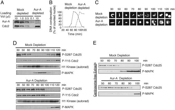
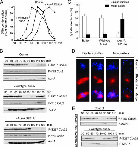
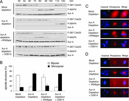
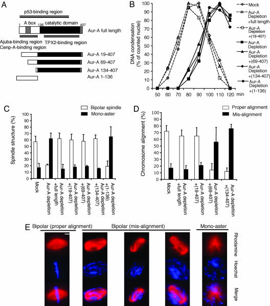
The kinase Aurora-A (Aur-A), which is enriched at centrosomes, is required for centrosome maturation and accurate chromosome segregation, and recent work implicates centrosomes as sites where the earliest activation of cyclin B1-cdc2 occurs. Here, we have used Xenopus egg extracts to investigate Aur-A's contribution to cell cycle progression and spindle morphology in the presence or absence of centrosomes. We find that addition of active Aur-A accelerates cdc2 activation and mitotic entry. Depletion of endogenous Aur-A or addition of inactive Aur-A, which lead to monopolar spindles, delays but does not block mitotic entry. These effects on timing and spindle structure do not require the presence of centrosomes or chromosomes. The catalytic domain alone of Aur-A is sufficient to restore spindle bipolarity; additional N-terminal sequences function in mitotic timing.


Conflict of interest statement

Conflict of interest statement: No conflicts declared.

Figures

Fig. 1.
Fig. 1.
Depletion of Aur-A delays mitotic entry, but does not block it. (A) Cycling extracts were prepared 30 min after egg activation and immunodepleted with two rounds of IgG (mock) or affinity-purified Aur-A antibody. Samples were analyzed by SDS/PAGE followed by blotting with Aur-A and cdc2 antibodies. (B) Mock or Aur-A-depleted cycling extracts were supplemented with demembranated sperm nuclei (500 per μl). Samples were stained with Hoechst dye 33342 and visualized by fluorescence microscopy. At least 100 chromosome structures at each time point were quantified. (C) Representative chromatin structures from B. (D) Samples were analyzed by SDS/PAGE followed by blotting with indicated antibodies. Cdc2 activity toward histone H1 was assayed by incorporation of γ-32P, followed by SDS/PAGE and autoradiography. (E) Cycling extracts lacking sperm nuclei were mock- or Aur-A-depleted. Samples were blotted with the indicated antibodies.
Fig. 2.
Fig. 2.
Overexpression of Aur-A accelerates the G2/M transition. Kinase-dead Aur-A delays mitotic entry and leads to monopolar spindles. (A) Recombinant wild-type or kinase-dead D281A Aur-A (100 ng/μl) or buffer was added to cycling extracts at 50 min. Samples were stained with Hoechst dye 33342. At least 100 nuclei were scored for each condition. (B) Samples in A were blotted with the indicated antibodies. (C) Cycling extracts were supplemented with nuclei and rhodamine-labeled tubulin. Wild-type or D281A Aur-A (100 ng/μl) or buffer was added at 50 min. Samples were stained with Hoechst dye 33342. The percentage of spindle structures was averaged from three independent experiments. At each time point, at least 100 spindle structures were counted. (D) Representative spindle structures are shown. (E) Cycling extracts lacking sperm nuclei were supplemented with recombinant wild-type Aur-A (100 ng/μl) or buffer at 50 min. Samples were blotted as above.
Fig. 3.
Fig. 3.
Adding back wild-type, but not kinase-dead form of Aur-A, to Aur-A-depleted cycling extracts corrects the timing of G2/M transition and rescues the bipolar spindle formation. (A) Mock or Aur-A-depleted extracts were supplemented with nuclei (500 per μl) and rhodamine-labeled tubulin. Wild-type Aur-A, kinase-dead D281A Aur-A, or buffer control was added to Aur-A-depleted cycling extracts at 30 min. Samples were analyzed as in Fig. 2. (B) Samples were taken during mitosis and stained with Hoechst dye 33342. The percentage of distinct spindle structures in mitosis was averaged from four independent experiments. At each time point, >100 spindle structures were counted. (C) Representative spindle structures are shown. (D) Sixty minutes after mitosis, samples were again taken and examined as above.
Fig. 4.
Fig. 4.
The Aur-A catalytic domain alone is sufficient to allow formation of bipolar spindle, but additional N-terminal sequences are required to restore the correct timing of mitotic entry and proper chromosome alignment. (A) Schematic of Aur-A proteins used in add-back experiments. (B) Mock- or Aur-A-depleted extracts were supplemented with nuclei (500 per μl) and rhodamine-labeled tubulin. Aur-A proteins or buffer were added to the Aur-A-depleted extract. Samples were analyzed as in Fig. 2. (C and D) The percentage of spindle structures and chromosome alignment was averaged from three independent experiments. At each time point, >100 spindle structures were counted. (E) Representative spindle structures. (Scale bar, 10 μm.)

References

    1. Blagden S. P., Glover D. M. Nat. Cell Biol. 2003;5:505–511. - PubMed
    1. Ducat D., Zheng Y. Exp. Cell Res. 2004;301:60–67. - PubMed
    1. Meraldi P., Honda R., Nigg E. A. Curr. Opin. Genet. Dev. 2004;14:29–36. - PubMed
    1. Varmark H. J. Cell Biochem. 2004;91:904–914. - PubMed
    1. Brittle A. L., Ohkura H. Curr. Biol. 2005;15:R880–R882. - PubMed

Publication types

Substances

LinkOut - more resources