. 2006 Apr 11;103(15):6037-42.
doi: 10.1073/pnas.0511324103. Epub 2006 Mar 31.

Severe urinary concentrating defect in renal collecting duct-selective AQP2 conditional-knockout mice

Affiliations

Severe urinary concentrating defect in renal collecting duct-selective AQP2 conditional-knockout mice

Aleksandra Rojek et al. Proc Natl Acad Sci U S A. .

Abstract

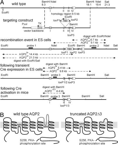
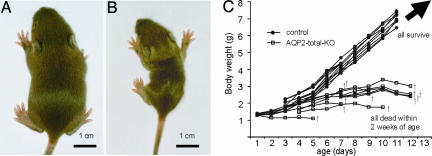
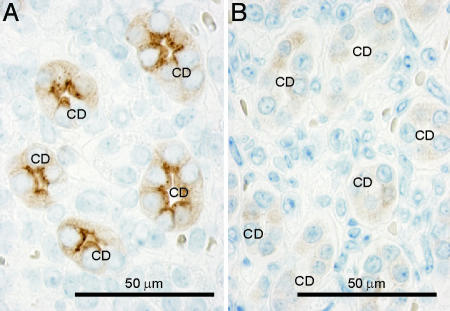
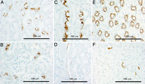
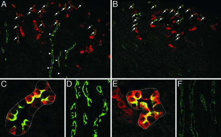
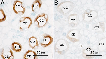
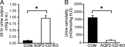
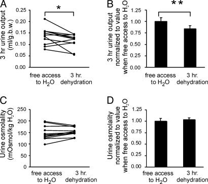
Aquaporin-2 (AQP2) is the predominant vasopressin-regulated water channel in kidney connecting tubule (CNT) and collecting duct (CD) and is essential for renal regulation of body water balance. However, the relative role of AQP2 to urinary concentration in the CNT and CD segments is unknown. To examine this directly, transgenic mice expressing AQP2 selectively in CNT but lacking AQP2 expression in CD (AQP2-CD-KO) and mice lacking AQP2 globally (AQP2-total-KO) were generated by exploiting the Cre/loxP technology. LoxP sites were inserted into AQP2 introns 2 and 3, and transgenic mice were bred with strains expressing Cre recombinase under the control of CD-specific Hoxb7- or global EIIa promoter. Mice lacking AQP2 globally died postnatally (days 5-12). AQP2-CD-KO mice were viable to adulthood and showed decreased body weight, 10-fold increased urine production (0.96 +/- 0.11 vs. 0.10 +/- 0.01 ml/g of body weight), and decreased urinary osmolality (170 +/- 19 vs. 1,630 +/- 135 milliosmoles/kg of H(2)O). Immunohistochemical staining of AQP2-CD-KO kidneys (n = 12) revealed sustained, strong AQP2 expression in CNT cells, whereas >95% of CD principal cells were completely AQP2-negative. Water deprivation for 3 hours caused only marginal decreased urine output (87 +/- 7% of levels when mice had free water access; P = 0.04) with no change in urine osmolality, revealing an absence of compensatory mechanisms. These results demonstrate that AQP2 in CNT is sufficient for postnatal survival and that AQP2 in CD is essential for regulation of body water balance and cannot be compensated for by other mechanisms.


Conflict of interest statement

Conflict of interest statement: No conflicts declared.

Figures

Fig. 1.
Fig. 1.
Targeted disruption of the mouse AQP2 gene. (A) Strategy for generating AQP2flx/flx mice. (B) Predicted peptide structures of wild-type AQP2 before (Left) and after (Right) deletion of exon 3. Deletion eliminates most of transmembrane domain 5, including the conserved NPA (Asn-Pro-Ala) motif in the channel-forming part of AQP2.
Fig. 2.
Fig. 2.
Growth retardation of AQP2-total-KO mice. A 10-day-old wild-type mouse (A) and a AQP2-total-KO mouse (B) from the same litter. The AQP2-total-KO mouse grew markedly less. (C) The growth of AQP2-total-KO and wild-type mice over time. All AQP2-total-KO mice died before day 13, whereas all controls survived.
Fig. 3.
Fig. 3.
Immunohistochemical labeling of AQP2 in control (A) and AQP2-total-KO (B) mice. In kidneys from control mice, strong apical labeling is associated with CD principal cells (CD). In contrast, weak and diffuse cytoplasmic labeling was observed in kidney CD principal cells in AQP2-total-KO mice, indicating the presence of low levels of truncated AQP2ΔE3 protein.
Fig. 4.
Fig. 4.
Immunohistochemical labeling of AQP2 in sections of kidneys from control (A, C, and E) and AQP2-CD-KO (B, D, and F) mice. A and B are from kidney cortex displaying CNTs. C and D are from kidney outer medulla displaying outer medullary CDs, and E and F are from kidney inner medulla displaying inner medullary CD. The CNT segment in the AQP2-CD-KO mice (B) expresses wild-type AQP2 protein.
Fig. 5.
Fig. 5.
Immunofluorescence double labeling of AQP2 (green) and a distal convoluted tubule and CNT marker calbindin D-28k (red) in sections of kidney cortex from control (A, C, and D) and AQP2-CD-KO (B, E, and F) mice. Strong AQP2 labeling was present in the CNT of both animals (A and B, arrows), whereas only the control mouse displayed strong AQP2 labeling in the calbindin D-28k-negative cortical CDs (A, arrowheads). (C and E) Higher magnification of CNT. (D and F) Higher magnification of CDs.
Fig. 6.
Fig. 6.
Immunohistochemical labeling of AQP2 phosphorylated at Ser-256 in sections of kidneys from control (A) and AQP2-CD-KO (B) mice. In kidneys from control mice, strong apical labeling was associated with CD principal cells (CD). In contrast, weak and diffuse cytoplasmic labeling was observed in CD in the AQP2-CD-KO mouse, indicating phosphorylation of the truncated AQP2ΔE3 protein.
Fig. 7.
Fig. 7.
AQP2-CD-KO mice had a markedly higher 24-h urine output compared with control mice (A) and markedly reduced urine osmolality compared with control mice (B). ∗, P < 0.05.
Fig. 8.
Fig. 8.
Changes in urine output (A and B) and urine osmolality (C and D) in response to free access to water or 3 h of complete water restriction of AQP2-CD-KO mice. The values were obtained in the same animals on consecutive days. Paired Student's t test reveals marginal, albeit statistically significant, reduction in urine output (∗, P = 0.04) but absence of significant differences in urine osmolality in response to 3 h of water restriction (B) (∗∗, P < 0.05).

Similar articles

Cited by

References

    1. Nielsen S., DiGiovanni S. R., Christensen E. I., Knepper M. A., Harris H. W. Proc. Natl. Acad. Sci. USA. 1993;90:11663–11667. - PMC - PubMed
    1. Nielsen S., Chou C. L., Marples D., Christensen E. I., Kishore B. K., Knepper M. A. Proc. Natl. Acad. Sci. USA. 1995;92:1013–1017. - PMC - PubMed
    1. Marples D., Knepper M. A., Christensen E. I., Nielsen S. Am. J. Physiol. 1995;269:C655–C664. - PubMed
    1. Sabolic I., Katsura T., Verbavatz J. M., Brown D. J. Membr. Biol. 1995;143:165–175. - PubMed
    1. Yamamoto T., Sasaki S., Fushimi K., Ishibashi K., Yaoita E., Kawasaki K., Marumo F., Kihara I. Am. J. Physiol. 1995;268:C1546–C1551. - PubMed

Publication types