

TGF beta-induced cartilage repair is maintained but fibrosis is blocked in the presence of Smad7
- PMID: 16584530
- PMCID: PMC1526625
- DOI: 10.1186/ar1931
TGF beta-induced cartilage repair is maintained but fibrosis is blocked in the presence of Smad7
Abstract
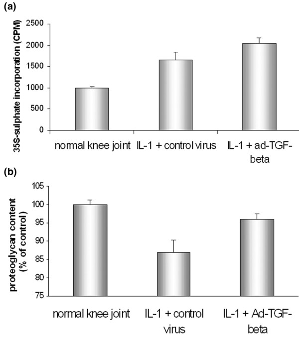
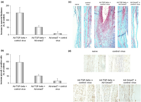
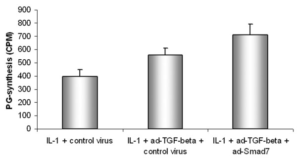
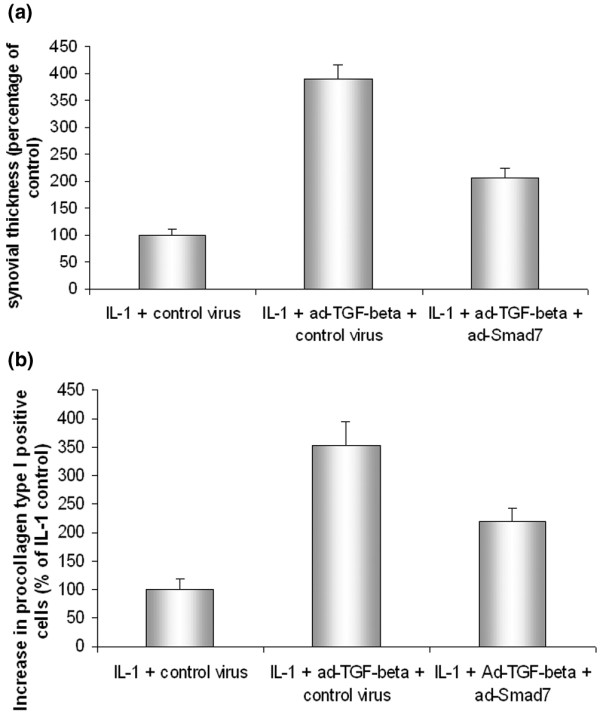
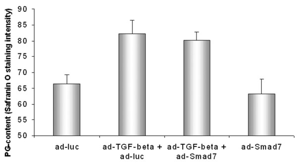
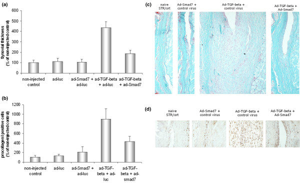
Cartilage damage in osteoarthritis (OA) is considered an imbalance between catabolic and anabolic factors, favoring the catabolic side. We assessed whether adenoviral overexpression of transforming growth factor-beta (TGFbeta) enhanced cartilage repair and whether TGFbeta-induced fibrosis was blocked by local expression of the intracellular TGFbeta inhibitor Smad7. We inflicted cartilage damage by injection of interleukin-1 (IL-1) into murine knee joints. After 2 days, we injected an adenovirus encoding TGFbeta. On day 4, we measured proteoglycan (PG) synthesis and content. To examine whether we could block TGFbeta-induced fibrosis and stimulate cartilage repair simultaneously, we injected Ad-TGFbeta and Ad-Smad7. This was performed both after IL-1-induced damage and in a model of primary OA. In addition to PG in cartilage, synovial fibrosis was measured by determining the synovial width and the number of procollagen I-expressing cells. Adenoviral overexpression of TGFbeta restored the IL-1-induced reduction in PG content and increased PG synthesis. TGFbeta-induced an elevation in PG content in cartilage of the OA model. TGFbeta-induced synovial fibrosis was strongly diminished by simultaneous synovial overexpression of Smad7 in the synovial lining. Of great interest, overexpression of Smad7 did not reduce the repair-stimulating effect of TGFbeta on cartilage. Adenoviral overexpression of TGFbeta stimulated repair of IL-1- and OA-damaged cartilage. TGFbeta-induced synovial fibrosis was blocked by locally inhibiting TGFbeta signaling in the synovial lining by simultaneously transfecting it with an adenovirus overexpressing Smad7.
Figures
Similar articles
-
Reduction of osteophyte formation and synovial thickening by adenoviral overexpression of transforming growth factor beta/bone morphogenetic protein inhibitors during experimental osteoarthritis.Arthritis Rheum. 2003 Dec;48(12):3442-51. doi: 10.1002/art.11328. Arthritis Rheum. 2003. PMID: 14673995
-
Resemblance of osteophytes in experimental osteoarthritis to transforming growth factor beta-induced osteophytes: limited role of bone morphogenetic protein in early osteoarthritic osteophyte formation.Arthritis Rheum. 2007 Dec;56(12):4065-73. doi: 10.1002/art.23034. Arthritis Rheum. 2007. PMID: 18050218
-
Stimulation of articular cartilage repair in established arthritis by local administration of transforming growth factor-beta into murine knee joints.Lab Invest. 1998 Feb;78(2):133-42. Lab Invest. 1998. PMID: 9484711
-
Gene therapy in musculoskeletal repair.Ann N Y Acad Sci. 2007 Nov;1117:310-27. doi: 10.1196/annals.1402.065. Ann N Y Acad Sci. 2007. PMID: 18056051 Review.
-
Cytokine targeting in osteoarthritis.Curr Drug Targets. 2007 Feb;8(2):283-92. doi: 10.2174/138945007779940179. Curr Drug Targets. 2007. PMID: 17305506 Review.
Cited by
-
Chondrocyte Hypertrophy in Osteoarthritis: Mechanistic Studies and Models for the Identification of New Therapeutic Strategies.Cells. 2022 Dec 13;11(24):4034. doi: 10.3390/cells11244034. Cells. 2022. PMID: 36552796 Free PMC article. Review.
-
The RNA-binding protein human antigen R controls global changes in gene expression during Schwann cell development.J Neurosci. 2012 Apr 4;32(14):4944-58. doi: 10.1523/JNEUROSCI.5868-11.2012. J Neurosci. 2012. PMID: 22492050 Free PMC article.
-
Gene expression profile in human induced pluripotent stem cells: Chondrogenic differentiation in vitro, part B.Mol Med Rep. 2017 May;15(5):2402-2414. doi: 10.3892/mmr.2017.6335. Epub 2017 Mar 16. Mol Med Rep. 2017. PMID: 28447733 Free PMC article.
-
Cell-free Stem Cell-Derived Extract Formulation for Regenerative Medicine Applications.Int J Mol Sci. 2020 Dec 9;21(24):9364. doi: 10.3390/ijms21249364. Int J Mol Sci. 2020. PMID: 33316880 Free PMC article.
-
Modulation of TGF-beta signaling by proinflammatory cytokines in articular chondrocytes.Osteoarthritis Cartilage. 2007 Dec;15(12):1367-77. doi: 10.1016/j.joca.2007.04.011. Epub 2007 Jun 29. Osteoarthritis Cartilage. 2007. PMID: 17604656 Free PMC article.
References
-
- Pedrozo HA, Schwartz Z, Gomez R, Ornoy A, Xin SW, Dallas SL, Bonewald LF, Dean DD, Boyan BD. Growth plate chondrocytes store latent transforming growth factor (TGF)-beta 1 in their matrix through latent TGF-beta 1 binding protein-1. J Cell Physiol. 1998;177:343–354. doi: 10.1002/(SICI)1097-4652(199811)177:2<343::AID-JCP16>3.0.CO;2-A. - DOI - PubMed
-
- Redini F, Min W, Demoor FM, Boittin M, Pujol JP. Differential expression of membrane-anchored proteoglycans in rabbit articular chondrocytes cultured in monolayers and in alginate beads. Effect of transforming growth factor-beta 1. Biochim Biophys Acta. 1997;1355:20–32. doi: 10.1016/S0167-4889(96)00115-2. - DOI - PubMed
Publication types
MeSH terms
Substances
LinkOut - more resources
Full Text Sources
