. 2006 Apr 11;103(15):6049-54.
doi: 10.1073/pnas.0600944103. Epub 2006 Apr 3.

The Arabidopsis homolog of trithorax, ATX1, binds phosphatidylinositol 5-phosphate, and the two regulate a common set of target genes

Affiliations

The Arabidopsis homolog of trithorax, ATX1, binds phosphatidylinositol 5-phosphate, and the two regulate a common set of target genes

Raul Alvarez-Venegas et al. Proc Natl Acad Sci U S A. .

Abstract

The Arabidopsis homolog of trithorax, ATX1, regulates numerous functions in Arabidopsis beyond the homeotic genes. Here, we identified genome-wide targets of ATX1 and showed that ATX1 is a receptor for a lipid messenger, phosphatidylinositol 5-phosphate, PI5P. PI5P negatively affects ATX1 activity, suggesting a regulatory pathway connecting lipid-signaling with nuclear functions. We propose a model to illustrate how plants may respond to stimuli (external or internal) that elevate cellular PI5P levels by altering expression of ATX1-controlled genes.


Conflict of interest statement

Conflict of interest statement: No conflicts declared.

Figures
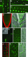
Fig. 1.
Fig. 1.
Distribution of ATX1-GFP in Arabidopsis root cells and tissues. (A–C) Root cells from transgenic plants expressing ATX1-GFP. ∗, position of the nuclei in cells where ATX1 was not nuclear. (D) In all cell types of recently emerged lateral root, ATX1-GFP is in the cytoplasm. Cell walls are counterstained with propidium iodide. (E) ATX1-GFP localization in the root cap of primary root. In peripheral columella cells (∗), ATX1 is dispersed throughout the cytoplasm, whereas, in the sloughing cells of the root cap, ATX1 is perinuclear (F and G). The nuclei are indicated by arrows. The image in G is Z- projection of eight optical sections, showing aggregates of ATX1 localized around the nuclei depicted in F. (H) Z-projection of five optical sections showing cytoplasmic and perinuclear localization of ATX1 in epidermal cells in the transition zone. (I) In epidermal cells of the elongation zone, ATX1 is detected along the plasma membrane (arrowheads); merged image of differential interference contrast microscopy (DIC) and Z-projection of four optical sections. (J) Nuclear localization of ATX1 in the epidermis within the transition zone. (K) Nuclear localization of ATX1 in the cortex (transition zone); Z-projection of nine optical sections. (L) Nuclear and membrane localization of ATX1 in the epidermis in the elongation zone; merged image of DIC and of a single optical section. EH, K, and L were taken from the same root (n = 26).
Fig. 2.
Fig. 2.
Structural models of PHDATX1, C1PKC, and FYVEEEA1. (A) Superposition of PHDATX peptide model (residues 599–648) on the structure of the Cys-2-domain (25). The ligand, DAG/PMA (black sticks), binds in a cleft formed between two loops. In the surface models of FYVEEEA1 (PDB 1HY), PHDATX (residues 606–667), and CYS2PKCD (PDB 1PTR), the basic patches are dark. The PI3P binding to FYVEEEA1 and PMA binding to CYS2PKCD are shown in black sticks. (B) Alignment of zinc-finger sequences belonging to different PHD families. Zinc coordination residues are shown in black-shaded white fonts. Light-shaded residues are involved in the positive patches at the PI3P-binding sites in VPS27, EEA1 (26), ING2 (17).
Fig. 3.
Fig. 3.
In vitro ligand binding by the protein-lipid overlay (PLO) assays. (A) Binding of recombinant GST-ATX1 to prespotted lipids. A key is shown to the right. (B) Affinity purified GST-ATX1 fusion protein reacted with membranes containing serially diluted ligands, as indicated: PMA, 4-α-phorbol-12-myristate-13-acetate; PI, phosphatidylinositol (PtdIns); PI3P, PtdIns(3)P; PI4P, PtdIns(4)P; PI5P, PtdIns(5)P; PI3,5P2, PtdIns(3,5)P2; P4,5P2, PtdIns(4,5)P2. Substrate concentrations in picomoles are shown. Bound proteins were revealed by GST-specific antibodies. (C) Duplicate control membrane reacted with the GST antibody. (D and E) Binding of recombinant GST-PHDING2 and GST-p40PX to membranes containing tested ligands. (F) PI5P-binding activity of GST-ATX1 and various deletion GST-fusion peptides. (G) Coomassie-stained SDS gels of total cellular proteins extracted from wild-type plants and from atx1 mutants. Western blot of the same extracts with the ATX1-specific antibody. Absence of the band in atx1 mutants confirms the specificity of the antibody. (H and I) Cellular ATX1 binds PI5P. Replicate membrane reacted with the ATX antibodies as a control for the specificity of the signal.
Fig. 4.
Fig. 4.
ATX1 in cells of Arabidopsis roots after treatment with PtdIns. (A) PI5P-induced changes in localization of ATX1-GFP. Time lapse experiments of roots treated with 1.5 μM of PI5P. Images taken 1 min, 50 min, and 95 min after drug application show Z-projections of 10, 9, and 10 optical sections, respectively. (A Top) The image on the right represents a merge of the (1 min) left image with the differential interference contrast microscopy (DIC) image. (Middle) After 95 min, most nuclei in the lower file of cells have almost completely lost nuclear signal. In contrast, exposure to PI4P for up to 2 h did not trigger loss of nuclear signal. (A Bottom) Right image is a merge of DIC with the image on the left. (B) Red particles inside root epidermis cells show presence of BODIPYR TMR-PI5P inside cells. Localization of ATX1-GFP protein is seen in green. Images shown in green and red channels are taken 30 min after treating roots in media supplied with 1.5 μM of red-tagged PI5P. (B Right) A merged image. Colocalization at the membrane (orange) may be seen along cell walls, particularly at the bulge of the emerging root hair (arrow). Inside the cytoplasm, PI5P is colocolized with ATX1 protein particles (some spots are shown by arrowheads). (C) Western blots of cytoplasm (Cy) and nuclear (Nu) proteins with ATX1 specific antibodies.
Fig. 5.
Fig. 5.
A model of a PI5P-ATX1 signaling pathway controlling Arabidopsis genes. Developmental and environmental factors influence the cellular concentration of PI5P. Higher levels of PI5P deactivate ATX1, which, in turn, represses transcription factors, a repressor in the case of some wall-modifying genes. Arrows, activation; T-shaped bars, repression events.

References

    1. Mazo A., Huang D.-H., Mozer B., Dawid I. Proc. Natl. Acad. Sci. USA. 1990;87:2112–2116. - PMC - PubMed
    1. Simon J. A., Tamkun J. W. Curr. Opin. Genet. Dev. 2002;12:210–218. - PubMed
    1. Schwartz-Sommer Z., Huijser P., Nacken W., Saedler H., Sommer H. Science. 1990;250:931–936. - PubMed
    1. Alvarez-Venegas R., Pien S., Sadder M., Witmer X., Grossniklaus U., Avramova Z. Curr. Biol. 2003;13:627–637. - PubMed
    1. Rea S., Eisenhaber F., O’Carroll D., Strahl B. D., Sun Z.-W., Schmid M., Opravil S., Mechtler K., Pontig C. P., Allis C. D., Jenuwein T. Nature. 2000;406:593–599. - PubMed

Publication types

MeSH terms

Substances