. 2006 Apr 11;103(15):5799-804.
doi: 10.1073/pnas.0510923103. Epub 2006 Apr 3.

Dominant-negative inhibition of the Axl receptor tyrosine kinase suppresses brain tumor cell growth and invasion and prolongs survival

Affiliations

Dominant-negative inhibition of the Axl receptor tyrosine kinase suppresses brain tumor cell growth and invasion and prolongs survival

Peter Vajkoczy et al. Proc Natl Acad Sci U S A. .

Abstract

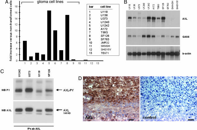
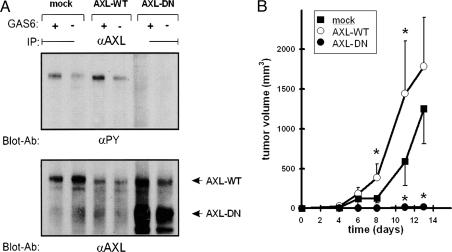
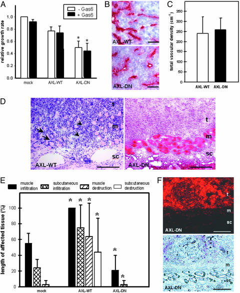
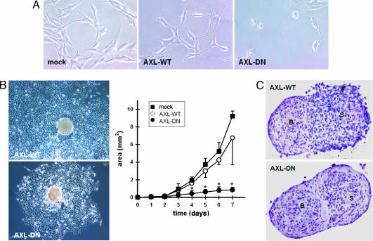
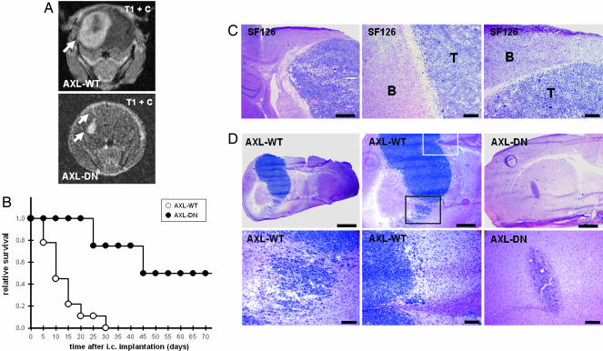
Malignant gliomas remain incurable brain tumors because of their diffuse-invasive growth. So far, the genetic and molecular events underlying gliomagenesis are poorly understood. In this study, we have identified the receptor tyrosine kinase Axl as a mediator of glioma growth and invasion. We demonstrate that Axl and its ligand Gas6 are overexpressed in human glioma cell lines and that Axl is activated under baseline conditions. Furthermore, Axl is expressed at high levels in human malignant glioma. Inhibition of Axl signaling by overexpression of a dominant-negative receptor mutant (AXL-DN) suppressed experimental gliomagenesis (growth inhibition >85%, P < 0.05) and resulted in long-term survival of mice after intracerebral glioma cell implantation when compared with Axl wild-type (AXL-WT) transfected tumor cells (survival times: AXL-WT, 10 days; AXL-DN, >72 days). A detailed analysis of the distinct hallmarks of glioma pathology, such as cell proliferation, migration, and invasion and tumor angiogenesis, revealed that inhibition of Axl signaling interfered with cell proliferation (inhibition 30% versus AXL-WT), glioma cell migration (inhibition 90% versus mock and AXL-WT, P < 0.05), and invasion (inhibition 62% and 79% versus mock and AXL-WT, respectively; P < 0.05). This study describes the identification, functional manipulation, in vitro and in vivo validation, and preclinical therapeutic inhibition of a target receptor tyrosine kinase mediating glioma growth and invasion. Our findings implicate Axl in gliomagenesis and validate it as a promising target for the development of approaches toward a therapy of these highly aggressive but, as yet, therapy-refractory, tumors.


Conflict of interest statement

Conflict of interest statement: P.V., P.K., and A.U. have filed a patent on the role of Axl in tumor biology. The patent has been licensed by the company U3, which pursues Axl as a potential target for antitumor therapy.

Figures

Fig. 1.
Fig. 1.
Expression of Axl in malignant glioma cells. (A) Relative expression level of Axl mRNA in human glioma cell lines (bars 1–9), neuroblastoma cell lines (bars 10–12), and a medulloblastoma cell line (bar 13) versus the nonneoplastic cell line HCN2 as assessed by cDNA array analysis. (B) Northern blot analysis of Axl and Gas6. Total RNA was isolated from human glioma cell lines and neuroblastoma cell lines. A β-actin probe was used as internal control. (C) Western blot analysis for Axl protein (WB AXL, Lower) and phosphotyrosine (WB PY, Upper) after immunoprecipitation of Axl protein. Cellular protein was isolated from four human glioma cell lines. (D) Immunohistochemical staining of Axl protein in a human glioblastoma multiforme specimen. Negative control staining was performed in the absence of the primary antibody. (Scale bar, 50 μm.)
Fig. 2.
Fig. 2.
Expression of a signaling-defective mutant form of Axl inhibits experimental tumor growth. (A) Western blot analysis of SF126 glioma cell clones expressing the control vector (mock), the wild-type form of Axl (AXL-WT), and a truncated dominant-negative mutant form of Axl (AXL-DN). Serum-depleted cells were left untreated (−) or treated with 200 μg/ml Gas6 (+). Lysates were blotted with anti-phosphotyrosine serum (Upper) or an antibody directed against the extracellular domain of human Axl (Lower). (B) Analysis of tumor volume for SF126 experimental cell clones. Tumor cells were implanted s.c. into nude mice. The mean ± SEM values are represented from eight animals per group. ∗, P < 0.05 vs. mock cells.
Fig. 3.
Fig. 3.
Inhibition of Axl signaling suppresses diffuse-invasive tumor growth. (A) Proliferation assay of transfected cell clones. Cells were left untreated (−Gas6) or treated with 200 μg/ml Gas6 (+Gas6). Analysis was performed after 48 h of culture. Growth rate is expressed in relation to unstimulated mock-transfected cells. Experiments were performed in triplicate. The mean ± SD values are represented. ∗, P < 0.05 versus mock. (B and C) Assessment of vessel density in AXL-WT and AXL-DN xenografts after s.c. implantation. Analysis of tumor blood vessel density and morphology was assessed by immunohistochemistry for CD31 (B) and quantitatively by intravital fluorescence videomicroscopy after tumor implantation into the skinfold chamber preparation (C). The mean ± SD values are represented from four animals per group. (Scale bars, 25 μm.) (D) Histomorphological images of AXL-WT tumors and AXL-DN tumors. Hematoxylin and eosin staining. Arrows indicate tumor cell invasion into the adjacent skin muscle layer resulting in displacement of the muscle tissue. Arrowheads indicate tumor cell invasion into the adjacent skin muscle layer resulting in destruction of the muscle tissue. t, tumor mass; m, skin muscle layer; sc, s.c. tissue. (Scale bars, 100 μm.) (E) Quantitative analysis of glioma cell invasion into adjacent tissue by mock, AXL-WT, and AXL-DN tumors. Linear analysis of the length of infiltrated and destroyed muscle and s.c. tissue is presented as percentage of the total cross-sectional length of tumor mass. The mean ± SD values are represented. ∗, P < 0.05 versus mock tumors. (F) Fluorescence microscopy alone (Upper) and in combination with phase contrast (Lower) of AXL-DN tumor. Tumor cells were labeled with DiI before implantation. (Scale bars, 100 μm.) All specimens (BF) were excised on day 21 after implantation into the dorsal skinfold chamber of nude mice. t, tumor mass; m, skin muscle layer; sc, s.c. tissue.
Fig. 4.
Fig. 4.
In vitro characterization reveals role for Axl in mediating glioma cell migration and invasion. (A) Analysis of morphology and in vitro behavior of cell clones under nonconfluent conditions. (B) Migration assay with multicellular aggregates of transfected cell clones. Migratory activity of tumor cells is illustrated for AXL-WT and AXL-DN cells at 72 h after plating (Left) and is analyzed quantitatively over 7 days (Right). Area of migration was analyzed planimetrically by means of an image analysis system. Experiments were performed in triplicate. The mean ± SD values are represented. ∗, P < 0.05 vs. mock-transfected cells. (C) Analysis of tumor cell invasion by 48-h confrontation of AXL-WT tumor cell spheroids (Upper) or AXL-DN tumor cell spheroids (Lower) with fetal rat brain cell aggregates. Note clear border between AXL-DN tumor cell spheroid and brain cell aggregate, whereas the AXL-WT spheroid has merged with the brain aggregate, indicating lack of invasiveness after inhibition of Axl signaling. B, brain cell aggregate; S, tumor spheroid. Experiments were performed in quadruplicate.
Fig. 5.
Fig. 5.
Inhibition of Axl signaling prolongs survival after orthotopic implantation. (A) MRI of intracerebral tumor growth on day 10 after stereotactic implantation of AXL-WT and AXL-DN cells into the brains of nude mice. T1-imaging sequence after injection of gadolinium-DTPA. (B) Survival curves for nude mice after intracerebral implantation of AXL-WT cells (n = 9 animals) and AXL-DN cells (n = 8 animals). Animals were killed as soon as they developed neurological deficits or lost >20% of their initial body weight. (C) Histomorphology of parental SF126 tumors after intracerebral implantation demonstrating the typical growth behavior of this cell line in the CNS. SF126 cells grow primarily as a solid mass and show only little invasion. Note the clear-cut border between the tumor (T) and brain tissue (B). Hematoxylin and eosin staining. [Scale bars, 500 μm (Left) and 100 μm (Center and Right).] (D) Histomorphology of AXL-WT tumors and AXL-DN tumors on day 10 after intracerebral implantation. Whereas AXL-WT tumors had developed to large, space-occupying lesions with diffuse tumor cell infiltration into adjacent brain tissue (Left and Center), AXL-DN tumor cells had formed only small and well demarcated tumors (Right). Squares depict areas of AXL-WT tumor cell invasion highlighted at higher magnification. AXL-WT tumor cells invaded either diffusely into the brain tissue (black square and Left Lower) or along white-matter tracts, such as the corpus callosum (white square and Center Lower). Hematoxylin and eosin staining. [Scale bars, 1 mm (Left Upper), 500 μm (Center and Right Upper), and 100 μm (Lower).]

Similar articles

Cited by

References

    1. DeAngelis L. M. N. Engl. J. Med. 2001;344:114–123. - PubMed
    1. Stupp R., Mason W. P., van den Bent M. J., Weller M., Fisher B., Taphoorn M. J., Belanger K., Brandes A. A., Marosi C., Bogdahn U., et al. N. Engl. J. Med. 2005;352:987–996. - PubMed
    1. Lefranc F., Brotchi J., Kiss R. J. Clin. Oncol. 2005;23:2411–2422. - PubMed
    1. Ullrich A., Schlessinger J. Cell. 1990;61:203–212. - PubMed
    1. Janssen J. W., Schulz A. S., Steenvoorden A. C., Schmidberger M., Strehl S., Ambros P. F., Bartram C. R. Oncogene. 1991;6:2113–2120. - PubMed

Publication types

MeSH terms