. 2006 Apr;188(8):2821-8.
doi: 10.1128/JB.188.8.2821-2828.2006.

Morphology of isolated Gli349, a leg protein responsible for Mycoplasma mobile gliding via glass binding, revealed by rotary shadowing electron microscopy

Affiliations

Morphology of isolated Gli349, a leg protein responsible for Mycoplasma mobile gliding via glass binding, revealed by rotary shadowing electron microscopy

Jun Adan-Kubo et al. J Bacteriol. 2006 Apr.

Abstract

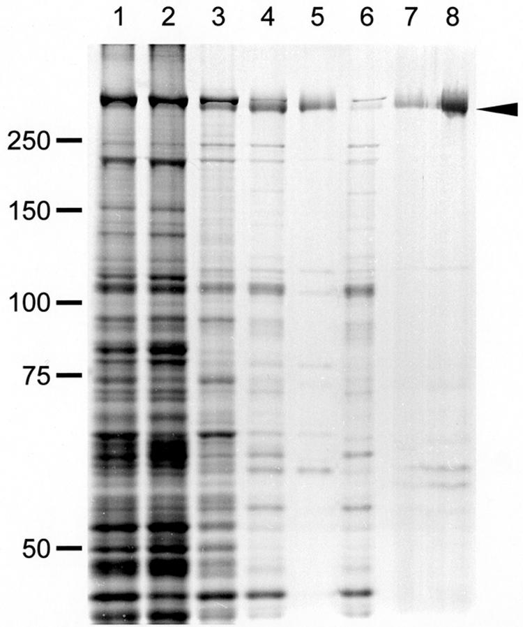
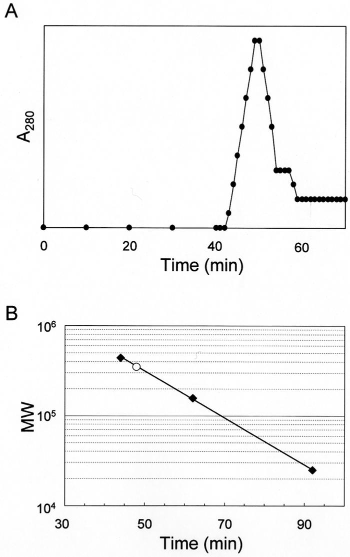
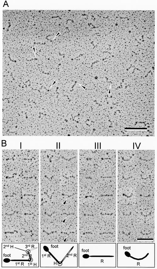
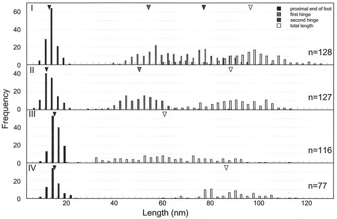
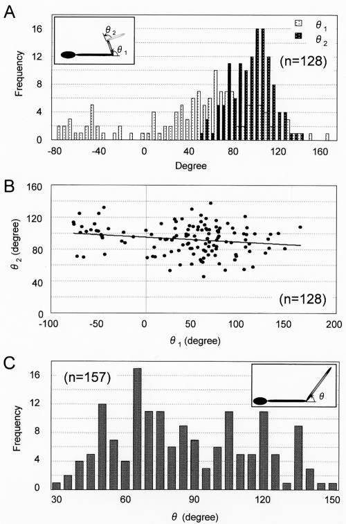
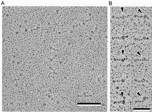
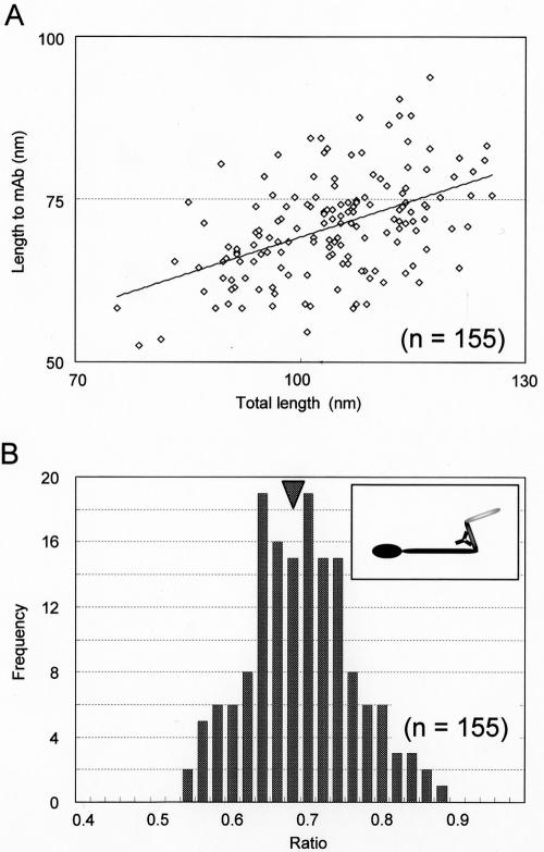
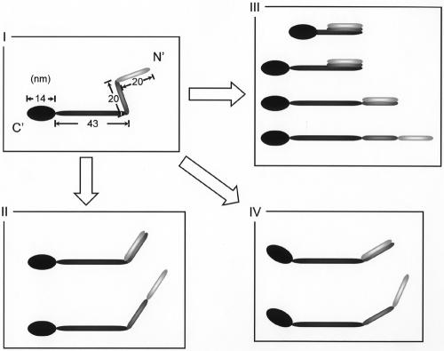
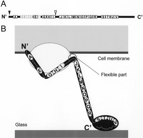
Several species of mycoplasmas rely on an unknown mechanism to glide across solid surfaces in the direction of a membrane protrusion at the cell pole. Our recent studies on the fastest species, Mycoplasma mobile, suggested that a 349-kDa protein, Gli349, localized at the base of the membrane protrusion called the neck, forms legs that stick out from the neck and propel the cell by repeatedly binding to and releasing from a solid surface, based on the energy of ATP hydrolysis. Here, the Gli349 protein was isolated from mycoplasma cells and its structure was analyzed. Gel filtration analysis showed that the isolated Gli349 protein is monomeric. Rotary shadowing electron microscopy revealed that the molecular structure resembles the symbol for an eighth note in music. It contains an oval foot 14 nm long in axis. From this foot extend three rods in tandem of 43, 20, and 20 nm, in that order. The hinge connecting the first and second rods is flexible, while the next hinge has a distinct preference in its angle, near 90 degrees. Molecular images revealed that a monoclonal antibody that can bind to the position at one-third of the total peptide length from the N terminus bound to a position two-thirds from the foot end, suggesting that the foot corresponds to the C-terminal region. The amino acid sequence was assigned to the molecular image, and the topology of the molecule in the gliding machinery is discussed.


Figures

FIG. 1.
FIG. 1.
Protein profiles of fractions in the Gli349 purification procedure. Gli349 protein was purified through four steps: (i) Triton X-100 treatment of cells and centrifugation, (ii) stepwise ammonium sulfate fractionation, (iii) precipitation of other proteins by a pH shift from 8.0 to 5.9, and (iv) Q-Sepharose (anion exchanger) column chromatography. The arrowhead shows the Gli349 protein band. Lane 1: whole-cell lysate. Lane 2: Triton-insoluble fraction of step 1. Lane 3: Triton-soluble fraction of step 1. Lane 4: precipitate of 35% saturation ammonium sulfate in step 2. Lane 5: supernatant of step 3. Lane 6: precipitate of step 3. Lane 7: fraction eluted at 0.15 M NaCl in step 4. Lane 8: 10-fold concentration of the fraction in lane 7. Each fraction was subjected to SDS-7.5% PAGE with a 3-mm lane width and stained by the reverse-staining method. Protein fractions derived from 0.2-, 0.5-, and 5-ml cultures were applied to lanes 1 to 6, 7, and 8, respectively. Molecular masses are indicated on the left in kilodaltons.
FIG. 2.
FIG. 2.
Gel filtration assay of isolated Gli349. (A) Elution pattern of Gli349 protein. (B) Molecular weight of native Gli349 protein. Molecular weight and retention time are presented on the y and x axes, respectively. Solid squares show size standards: ferritin (440 kDa), aldolase (158 kDa), and chymotrypsinogen (25 kDa). The retention time and calculated molecular weight (MW) of Gli349 are presented by the open circle.
FIG. 3.
FIG. 3.
Rotary-shadowed EM of Gli349 molecules. (A) Image of a field 1,354 nm wide and 1,122 nm high. Arrowheads indicate spherical and thick parts, called feet. Bar, 200 nm. (B) Isolated images and classification into four types. The molecular images were classified into types I, II, III, and IV, featuring two bends, one bend, linear form with no bend, and curved form with no bend, respectively. The images are aligned so that the feet are on the left side. Ten molecular images are presented for each type indicated at the top of each column. A distinct bend was observed in type II images, as marked by triangles, but not in type IV images. The schematics below each image present each part of the image, as the first rod (R), first hinge (H), and so on. Bar, 100 nm.
FIG. 4.
FIG. 4.
Positions of features from distal ends of the feet in Gli349 images. The molecule types are indicated on the left in each histogram. Triangles indicate averages.
FIG. 5.
FIG. 5.
Angles around hinges of type I and II Gli349 images. (A) Distribution of hinge angles θ1 and θ2 in type I images. The inset shows a schematic of the type I image. (B) Relationship between two angles in type I images. The solid line shows the linearized approximation. (C) Distribution of angle θ in type II images. The inset shows a schematic of the type II image.
FIG. 6.
FIG. 6.
Rotary-shadowed Gli349 molecules decorated by antibody MAb7. (A) Image 900 nm wide and 900 nm high. Bar, 200 nm. (B) Isolated images of Gli349 molecules coated with MAb7. Foot ends are directed to the left. Arrowheads indicate MAb7 bound to Gli349. Bar, 100 nm.
FIG. 7.
FIG. 7.
Positions of MAb7 on molecular images of Gli349. (A) Relationship between positions of MAb7 on Gli349 images and total length. The solid line shows a linearized approximation. (B) Distribution of MAb7 positions on Gli349 images. The central positions of MAb7 are presented as a ratios of the total length of the molecule. The average was 0.682 (n = 155), as indicated by the arrowhead. The inset shows a schematic illustration of a type I molecule coated with MAb7 at the average position.
FIG. 8.
FIG. 8.
Schematic of relationships among the four types of molecular images. Type I images retain the most features.
FIG. 9.
FIG. 9.
Schematic of the Gli349 molecule. (A) Amino acid sequence. Solid and broken boxes indicate repeats found in Gli349 and positions corresponding to those found only in the ortholog produced by M. pulmonis, MYPU2110, respectively. The names of repeats A to V (13) are indicated on the boxes. The predicted transmembrane segment and epitope of MAb7 are indicated by solid and open triangles, respectively. (B) Amino acid sequence and topology in the cell membrane. The cytoplasm and membrane of a mycoplasmal cell are shown by the meshed area and the thick line, respectively. The glass surface is shown as a gray line. The Gli349 molecule is presented as a rod sticking out of the cell and is supported by other protein components, represented by the gray ellipse. The repeat sequences and gap regions are represented by small ellipses and gray lines, respectively.

References

    1. Aluotto, B. B., R. G. Wittler, C. O. Williams, and J. E. Faber. 1970. Standardized bacteriologic techniques for the characterization of mycoplasma species. Int. J. Syst. Bacteriol. 20:35-58.
    1. Arata, T. 1998. Electron microscopic observation of monomeric actin attached to a myosin head. J. Struct. Biol. 123:8-16. - PubMed
    1. Bredt, W. 1979. Motility, p. 141-145. In M. F. Barile, S. Razin, J. G. Tully, and R. F. Whitcomb (ed.), The mycoplasmas, vol. 1. Academic Press, New York, N.Y.
    1. Charon, N. W. 2005. Mycoplasma takes a walk. Proc. Natl. Acad. Sci. USA 102:13713-13714. - PMC - PubMed
    1. Fernandez-Patron, C., L. Castellanos-Serra, and P. Rodriguez. 1992. Reverse staining of sodium dodecyl sulfate polyacrylamide gels by imidazole-zinc salts: sensitive detection of unmodified proteins. BioTechniques 12:564-573. - PubMed

Publication types

MeSH terms

LinkOut - more resources