. 2006 Apr;188(8):2845-55.
doi: 10.1128/JB.188.8.2845-2855.2006.

Transcriptional heat shock response in the smallest known self-replicating cell, Mycoplasma genitalium

Affiliations

Transcriptional heat shock response in the smallest known self-replicating cell, Mycoplasma genitalium

Oxana Musatovova et al. J Bacteriol. 2006 Apr.

Abstract

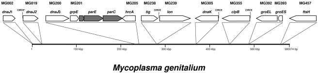
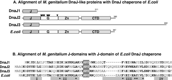
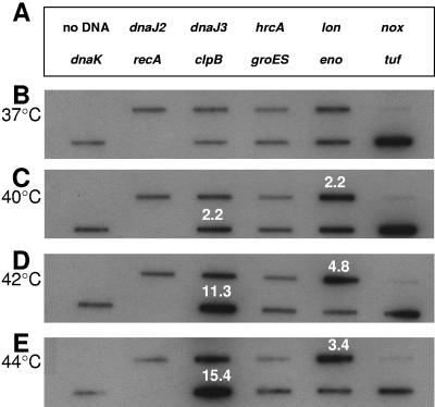
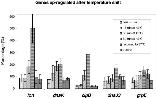
Mycoplasma genitalium is a human bacterial pathogen linked to urethritis and other sexually transmitted diseases as well as respiratory and joint pathologies. Though its complete genome sequence is available, little is understood about the regulation of gene expression in this smallest known, self-replicating cell, as its genome lacks orthologues for most of the conventional bacterial regulators. Still, the transcriptional repressor HrcA (heat regulation at CIRCE [controlling inverted repeat of chaperone expression]) is predicted in the M. genitalium genome as well as three copies of its corresponding regulatory sequence CIRCE. We investigated the transcriptional response of M. genitalium to elevated temperatures and detected the differential induction of four hsp genes. Three of the up-regulated genes, which encode DnaK, ClpB, and Lon, possess CIRCE within their promoter regions, suggesting that the HrcA-CIRCE regulatory mechanism is functional. Additionally, one of three DnaJ-encoding genes was up-regulated, even though no known regulatory sequences were found in the promoter region. Transcript levels returned to control values after 1 h of incubation at 37 degrees C, reinforcing the transient nature of the heat shock transcriptional response. Interestingly, neither of the groESL operon genes, which encode the GroEL chaperone and its cochaperone GroES, responded to heat shock. These data suggest that M. genitalium selectively regulates a limited number of genes in response to heat shock.


Figures

FIG. 1.
FIG. 1.
Schematic diagram of the position and organization of heat-stress related genes in the M. genitalium genome. Presumed heat shock-related genes are pictured as clear open arrows, and their orientations represent the corresponding coding strand. Closed arrows show genes separating hrcA from dnaJ3 and grpE. Genes are designated by their locus numbers and names as listed at www.tigr.org. Also, their position on the chromosome is illustrated. Copies of DnaJ-like protein coding genes are numbered 1 through 3 for convenience. All copies of CIRCE regulatory elements are shown; the least-conserved copy is labeled with asterisk.
FIG. 2.
FIG. 2.
Comparison of M. genitalium DnaJ-like proteins with E. coli chaperone DnaJ. (A) Structural comparison of M. genitalium DnaJ-like proteins DnaJ1, DnaJ2, and DnaJ3 with chaperone DnaJ of E. coli. The J domains (J), glycine-rich region (G; arrow heads indicate glycine residues), zinc-finger domain (Zn), and carboxy-terminal domain (CTD) are illustrated. (B) Alignment of J domains. Gray boxes illustrate positions of predicted helices (I to IV). Asterisks show hydrophobic residues that are required for the stabilization of helices. The conserved motif HPD in the loop between helices II and III is indicated. Bold letters represent basic residues.
FIG. 3.
FIG. 3.
Transcriptional analysis of dnaJ2. (A) Reverse transcription PCR was performed on total RNA isolated from M. genitalium cells, which generated product (RT+) as visualized on 1.2% agarose. To prove the absence of DNA in RNA preparations, a parallel reaction was run without reverse transcriptase (RT−). Genomic DNA was used to demonstrate the specificity of primers by amplification of a single 535-bp product. A 100-bp DNA ladder (Gibco BRL) was run alongside. (B) Identification of dnaJ2 TSP. PE and DNA sequencing products (lanes A, C, G, and T) were generated by end-labeled primer MG019PE and separated on 6% urea-polyacrylamide sequencing gel. The promoter region of dnaJ2 is at bottom, with the shaded area representing the putative −10 region, the asterisk designating the transcriptional start (bold C), and the bold ATG indicating the putative translational start.
FIG. 4.
FIG. 4.
Transcriptional analysis of M. genitalium heat shock genes. (A) Promoter regions and transcriptional start points. Asterisks designate transcriptional starts (bold letters) that were determined by PE. Shaded areas of sequences indicate putative −10 regions. The bold ATG (far right) in each sequence indicates the putative translational start. Underlined sequences (solid lines) represent CIRCE elements. IRs of CIRCE elements are shown in bold. The dashed underline indicates a CIRCE-like sequence upstream of dnaJ2 (MG019), and only conserved nucleotides of the IR are bolded. The CIRCE sequence is indicated above the dnaK promoter region. (B) Analysis of transcription by RT-PCR. Gene schematics with identified transcriptional starts are presented. Thin lines represent regions that are amplified to confirm the transcription of individual genes. Bold lines represent RT-PCR products indicating the cotranscription of corresponding genes. Numbers represent the size of RT-PCR-amplified products separated on 1% agarose (RT+ lanes), alongside corresponding reactions without reverse transcriptase (RT− lanes) and PCRs using M. genitalium genomic DNA (DNA lanes) to demonstrate specificity of primers.
FIG. 5.
FIG. 5.
Autoradiograph of DNA blot arrays for selected M. genitalium genes. (A) Designations of gene-specific PCR products blotted onto Zeta probe membrane. Membranes were hybridized with radiolabeled cDNAs that were generated from M. genitalium incubated at 37 (B), 40 (C), 42 (D), and 44°C (E) for 30 min. Synthesis of cDNA and other procedures are described in Materials and Methods. Numerical values indicate an increase in the amount of transcripts after temperature shift (value [n-fold] of transcript amount at 37°C).
FIG. 6.
FIG. 6.
Histogram of up-regulated heat shock genes of M. genitalium incubated at 42°C for different times. Housekeeping gene eno was used as the normalizer, and values represent each mRNA as the percentage of eno transcript. Data are presented as means ± standard deviations (error bars) (three experiments, each performed in triplicate). “Returned to 37°C” represents cells that were subjected to 42°C for 60 min and returned to 37°C for 60 min before RNA isolation. “Control” represents cells that were incubated at 37°C for 120 min before RNA isolation.
FIG. 7.
FIG. 7.
Interaction between HrcA- and CIRCE-containing promoter regions. DNA probes for M. genitalium dnaK and dnaJ2 and C. trachomatis dnaK promoter regions (lanes 1) were amplified and interacted with chlamydial HrcA protein (lanes 2). The presence of unlabeled competitive probe in reaction mix (lanes 3) confirmed specificity of interaction.

References

    1. Bailey, C. C., and K. F. Bott. 1994. An unusual gene containing a dnaJ N-terminal box flanks the putative origin of replication of Mycoplasma genitalium. J. Bacteriol. 176:5814-5819. - PMC - PubMed
    1. Bang, H., A. Pecht, G. Raddatz, T. Scior, W. Solbach, K. Brune, and A. Pahl. 2000. Prolyl isomerases in a minimal cell. Catalysis of protein folding by trigger factor from Mycoplasma genitalium. Eur. J. Biochem. 267:3270-3280. - PubMed
    1. Baseman, J. B., M. Cagle, J. E. Korte, C. Herrera, W. G. Rasmussen, J. G. Baseman, R. Shain, and J. M. Piper. 2004. Diagnostic assessment of Mycoplasma genitalium in culture-positive women. J. Clin. Microbiol. 42:203-211. - PMC - PubMed
    1. Baseman, J. B., S. F. Dallo, J. G. Tully, and D. L. Rose. 1988. Isolation and characterization of Mycoplasma genitalium strains from the human respiratory tract. J. Clin. Microbiol. 26:2266-2269. - PMC - PubMed
    1. Blaylock, M. W., O. Musatovova, J. G. Baseman, and J. B. Baseman. 2004. Determination of infectious load of Mycoplasma genitalium in clinical samples of human vaginal cells. J. Clin. Microbiol. 42:746-752. - PMC - PubMed

Publication types

MeSH terms