Functional domains of the Bacillus subtilis transcription factor AraR and identification of amino acids important for nucleoprotein complex assembly and effector binding
- PMID: 16585763
- PMCID: PMC1446991
- DOI: 10.1128/JB.188.8.3024-3036.2006
Functional domains of the Bacillus subtilis transcription factor AraR and identification of amino acids important for nucleoprotein complex assembly and effector binding
Abstract
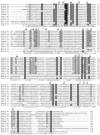
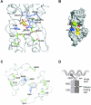
The Bacillus subtilis AraR transcription factor represses at least 13 genes required for the extracellular degradation of arabinose-containing polysaccharides, transport of arabinose, arabinose oligomers, xylose, and galactose, intracellular degradation of arabinose oligomers, and further catabolism of this sugar. AraR exhibits a chimeric organization comprising a small N-terminal DNA-binding domain that contains a winged helix-turn-helix motif similar to that seen with the GntR family and a larger C-terminal domain homologous to that of the LacI/GalR family. Here, a model for AraR was derived based on the known crystal structures of the FadR and PurR regulators from Escherichia coli. We have used random mutagenesis, deletion, and construction of chimeric LexA-AraR fusion proteins to map the functional domains of AraR required for DNA binding, dimerization, and effector binding. Moreover, predictions for the functional role of specific residues were tested by site-directed mutagenesis. In vivo analysis identified particular amino acids required for dimer assembly, formation of the nucleoprotein complex, and composition of the sugar-binding cleft. This work presents a structural framework for the function of AraR and provides insight into the mechanistic mode of action of this modular repressor.
Figures


Similar articles
-
Probing key DNA contacts in AraR-mediated transcriptional repression of the Bacillus subtilis arabinose regulon.Nucleic Acids Res. 2007;35(14):4755-66. doi: 10.1093/nar/gkm509. Epub 2007 Jul 7. Nucleic Acids Res. 2007. PMID: 17617643 Free PMC article.
-
Negative regulation of L-arabinose metabolism in Bacillus subtilis: characterization of the araR (araC) gene.J Bacteriol. 1997 Mar;179(5):1598-608. doi: 10.1128/jb.179.5.1598-1608.1997. J Bacteriol. 1997. PMID: 9045819 Free PMC article.
-
Structure of the effector-binding domain of the arabinose repressor AraR from Bacillus subtilis.Acta Crystallogr D Biol Crystallogr. 2012 Feb;68(Pt 2):176-85. doi: 10.1107/S090744491105414X. Epub 2012 Jan 17. Acta Crystallogr D Biol Crystallogr. 2012. PMID: 22281747 Free PMC article.
-
Allosteric control of transcription in GntR family of transcription regulators: A structural overview.IUBMB Life. 2015 Jul;67(7):556-63. doi: 10.1002/iub.1401. Epub 2015 Jul 14. IUBMB Life. 2015. PMID: 26172911 Review.
-
Ribbon-helix-helix transcription factors: variations on a theme.Nat Rev Microbiol. 2007 Sep;5(9):710-20. doi: 10.1038/nrmicro1717. Nat Rev Microbiol. 2007. PMID: 17676053 Review.
Cited by
-
Autoregulator protein PhaR for biosynthesis of polyhydroxybutyrate [P(3HB)] possibly has two separate domains that bind to the target DNA and P(3HB): Functional mapping of amino acid residues responsible for DNA binding.J Bacteriol. 2007 Feb;189(3):1118-27. doi: 10.1128/JB.01550-06. Epub 2006 Nov 22. J Bacteriol. 2007. PMID: 17122335 Free PMC article.
-
Functional Analysis of the Citrate Activator CitO from Enterococcus faecalis Implicates a Divalent Metal in Ligand Binding.Front Microbiol. 2016 Feb 9;7:101. doi: 10.3389/fmicb.2016.00101. eCollection 2016. Front Microbiol. 2016. PMID: 26903980 Free PMC article.
-
Structural and functional characterization of the LldR from Corynebacterium glutamicum: a transcriptional repressor involved in L-lactate and sugar utilization.Nucleic Acids Res. 2008 Dec;36(22):7110-23. doi: 10.1093/nar/gkn827. Epub 2008 Nov 6. Nucleic Acids Res. 2008. PMID: 18988622 Free PMC article.
-
Interaction of the GntR-family transcription factor Sll1961 with thioredoxin in the cyanobacterium Synechocystis sp. PCC 6803.Sci Rep. 2018 Apr 27;8(1):6666. doi: 10.1038/s41598-018-25077-5. Sci Rep. 2018. PMID: 29703909 Free PMC article.
-
Molecular and Functional Insights into the Regulation of d-Galactonate Metabolism by the Transcriptional Regulator DgoR in Escherichia coli.J Bacteriol. 2019 Jan 28;201(4):e00281-18. doi: 10.1128/JB.00281-18. Print 2019 Feb 15. J Bacteriol. 2019. PMID: 30455279 Free PMC article.
References
-
- Bell, C. E., and M. Lewis. 2000. A closer view of the conformation of the Lac repressor bound to operator. Nat. Struct. Biol. 7:209-214. - PubMed
-
- Bell, C. E., and M. Lewis. 2001. The Lac repressor: a second generation of structural and functional studies. Curr. Opin. Struct. Biol. 11:19-25. - PubMed
-
- Bjorkman, A. J., R. A. Binnie, H. Zhang, L. B. Cole, M. A. Hermodson, and S. L. Mowbray. 1994. Probing protein-protein interactions. The ribose-binding protein in bacterial transport and chemotaxis. J. Biol. Chem. 269:30206-30211. - PubMed
-
- Brennan, R. G. 1993. The winged-helix DNA-binding motif: another helix-turn-helix takeoff. Cell 74:773-776. - PubMed
-
- Daines, D. A., M. Granger-Schnarr, M. Dimitrova, and R. P. Silver. 2002. Use of LexA-based system to identify protein-protein interactions in vivo. Methods Enzymol. 358:153-161. - PubMed
Publication types
MeSH terms
Substances
LinkOut - more resources
Full Text Sources
Other Literature Sources
Molecular Biology Databases
Miscellaneous

