

doi: 10.1128/JB.188.8.3110-3115.2006.
EspF of enteropathogenic Escherichia coli binds sorting nexin 9
Affiliations
- PMID: 16585770
- PMCID: PMC1447016
- DOI: 10.1128/JB.188.8.3110-3115.2006
Item in Clipboard
EspF of enteropathogenic Escherichia coli binds sorting nexin 9
J Bacteriol.
2006 Apr.
Abstract
EspF of enteropathogenic Escherichia coli targets mitochondria and subverts a number of cellular functions. EspF consists of six putative Src homology 3 (SH3) domain binding motifs. In this study we identified sorting nexin 9 (SNX9) as a host cell EspF binding partner protein, which binds EspF via its amino-terminal SH3 region. Coimmunoprecipitation and confocal microscopy showed specific EspF-SNX9 interaction and non-mitochondrial protein colocalization in infected epithelial cells.
Figures

A. Identification of SNX9 as an EspF target protein. Yeast containing pICC175 (encoding EspF) and pICC347 (encoding SNX9) demonstrated a ca. 35-fold increase in β-galactosidase activity compared to single-plasmid-bearing strains. Coexpression of EspF and SNX9ΔSH3 did not activate the β-galactosidase reporter gene. B. Schematic domain organization of SNX9 (not to scale).

EspF-SNX9214 and EspF-SNX9FL protein interactions. Extracts of E. coli cell strains overexpressing MBP-SNX9214 (lanes 1), MBP-SNX9ΔSH3 (lanes 2), MBP (lanes 3), and GST-SNX9FL (lanes 4) were separated on sodium dodecyl sulfate-polyacrylamide gels (A), transferred to nitrocellulose membranes, and probed with anti-MBP (B) or overlaid with His-EspF (C). Similar expression levels of MBP and GST protein derivatives are seen (A and B), but EspF bound specifically to MBP-SNX9214 and GST-SNX9FL (C). Numbers at left are molecular masses in kilodaltons.
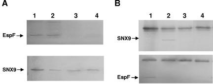
A. Detection of EspF and SNX9 in membrane and cytoplasmic fractions of HAC-7 cells infected with wt EPEC (lanes 1 and 2) and EPECΔespF (lanes 3 and 4). EspF was detected in both fractions only after infection with wt EPEC. SNX9 was detected in both cell fractions regardless of the EPEC strain used for infection. B. (Upper panel) IP with anti-EspF and detection with anti-SNX9, using membrane (lanes 2 and 4) and cytoplasmic (lanes 1 and 3) extracts of HAC-7 cells infected with wt EPEC (lanes 1 and 2) or EPECΔespF (lanes 3 and 4). SNX9 was specifically coimmunoprecipitated with EspF from the membrane extract infected with wt EPEC (lane 2) but not with EPECΔespF. (Lower panel) IP with anti-SNX9 and detection with anti-EspF, using membrane (lanes 1 and 3) and cytoplasmic (lanes 2 and 4) extracts of HAC-7 cells infected with wt EPEC (lanes 1 and 2) or EPECΔespF (lanes 3 and 4). EspF was specifically coimmunoprecipitated with SNX9 from the membrane extract infected with wt EPEC (lane 1) but not with EPECΔespF.

(Top) Translocation of EspF into HeLa cells. Confocal images show HeLa cells infected for 10, 30, and 120 min with primed cultures of wt E2348/69 and for 120 min with an espF mutant. Cells were stained for EspF and cell actin. EspF was detected beneath bacterial microcolonies (sites of actin accretion, arrows) after 10 min, but by 30 min EspF staining was also seen in adjacent filamentous organelles identified as mitochondria; after 2 h staining was predominantly in mitochondria. The espF mutant induced actin accretion but did not translocate EspF. (Bottom) Colocalization of SNX9 and EspF. Confocal images show uninfected HeLa cells and cells infected for 1 h with E2348/69 and stained for SNX9 and EspF. A punctate distribution of SNX9 was seen in uninfected cells, but following EPEC infection SNX9 became concentrated beneath bacterial colonies and colocalized with EspF (overlay, yellow); EspF that had migrated to adjacent mitochondria did not colocalize with SNX9 (overlay, red).

Localization of clathrin. Confocal images show uninfected HeLa cells and cells infected for 1 h with E2348/69 and stained for clathrin and cell actin. Clathrin was seen throughout the cell, but concentrations were frequently seen in a perinuclear region of the cell. This distribution was unchanged following EPEC infection, and there was no clathrin accumulation beneath sites of bacterial adhesion (arrow). Bar = 5 μm.
Similar articles
-
EPEC effector EspF promotes Crumbs3 endocytosis and disrupts epithelial cell polarity.Cell Microbiol. 2017 Nov;19(11):10.1111/cmi.12757. doi: 10.1111/cmi.12757. Epub 2017 Jul 27. Cell Microbiol. 2017. PMID: 28618099 Free PMC article.
-
The type III effector EspF coordinates membrane trafficking by the spatiotemporal activation of two eukaryotic signaling pathways.J Cell Biol. 2007 Sep 24;178(7):1265-78. doi: 10.1083/jcb.200705021. J Cell Biol. 2007. PMID: 17893247 Free PMC article.
-
E. coli secreted protein F promotes EPEC invasion of intestinal epithelial cells via an SNX9-dependent mechanism.Cell Microbiol. 2010 Jul;12(7):919-29. doi: 10.1111/j.1462-5822.2010.01440.x. Epub 2010 Jan 20. Cell Microbiol. 2010. PMID: 20088948 Free PMC article.
-
Comparative analysis of EspF variants in inhibition of Escherichia coli phagocytosis by macrophages and inhibition of E. coli translocation through human- and bovine-derived M cells.Infect Immun. 2011 Nov;79(11):4716-29. doi: 10.1128/IAI.00023-11. Epub 2011 Aug 29. Infect Immun. 2011. PMID: 21875965 Free PMC article.
-
Sorting out the cellular functions of sorting nexins.Nat Rev Mol Cell Biol. 2002 Dec;3(12):919-31. doi: 10.1038/nrm974. Nat Rev Mol Cell Biol. 2002. PMID: 12461558 Review.
Cited by
-
Exploitation of eukaryotic subcellular targeting mechanisms by bacterial effectors.Nat Rev Microbiol. 2013 May;11(5):316-26. doi: 10.1038/nrmicro3009. Nat Rev Microbiol. 2013. PMID: 23588250 Free PMC article. Review.
-
Mitotic Arrest-Deficient 2 Like 2 (MAD2L2) Interacts with Escherichia coli Effector Protein EspF.Life (Basel). 2021 Sep 15;11(9):971. doi: 10.3390/life11090971. Life (Basel). 2021. PMID: 34575120 Free PMC article.
-
EPEC effector EspF promotes Crumbs3 endocytosis and disrupts epithelial cell polarity.Cell Microbiol. 2017 Nov;19(11):10.1111/cmi.12757. doi: 10.1111/cmi.12757. Epub 2017 Jul 27. Cell Microbiol. 2017. PMID: 28618099 Free PMC article.
-
EspJ of enteropathogenic and enterohaemorrhagic Escherichia coli inhibits opsono-phagocytosis.Cell Microbiol. 2008 May;10(5):1104-15. doi: 10.1111/j.1462-5822.2007.01112.x. Epub 2008 Jan 14. Cell Microbiol. 2008. PMID: 18201246 Free PMC article.
-
Actin cytoskeleton manipulation by effector proteins secreted by diarrheagenic Escherichia coli pathotypes.Biomed Res Int. 2013;2013:374395. doi: 10.1155/2013/374395. Epub 2012 Dec 30. Biomed Res Int. 2013. PMID: 23509714 Free PMC article. Review.
References
-
- Crane, J. K., B. P. McNamara, and M. S. Donnenberg. 2001. Role of EspF in host cell death induced by enteropathogenic Escherichia coli. Cell. Microbiol. 3:197-211. - PubMed
-
- Creasey, E. A., R. M. Delahay, S. J. Daniell, and G. Frankel. 2003. Yeast two-hybrid system survey of interactions between LEE-encoded proteins of enteropathogenic Escherichia coli. Microbiology 149:2093-2106. - PubMed
-
- Frankel, G., A. D. Phillips, L. R. Trabulsi, S. Knutton, G. Dougan, and S. E. Matthews. 2001. Intimin and the host cell—is it bound to end in Tir(s)? Trends Microbiol. 9:214-218. - PubMed
Publication types
MeSH terms
Substances
Grants and funding
LinkOut - more resources
Full Text Sources
Other Literature Sources
Molecular Biology Databases
Miscellaneous
