. 2006 Apr;116(4):940-52.
doi: 10.1172/JCI22689.

Neotendon formation induced by manipulation of the Smad8 signalling pathway in mesenchymal stem cells

Affiliations

Neotendon formation induced by manipulation of the Smad8 signalling pathway in mesenchymal stem cells

Andrea Hoffmann et al. J Clin Invest. 2006 Apr.

Abstract

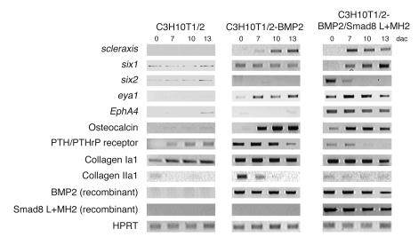
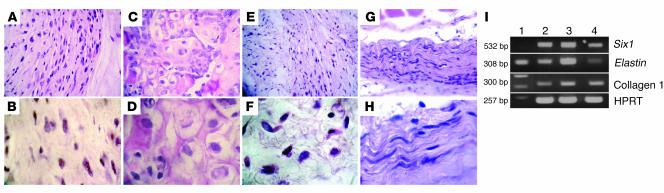
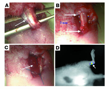
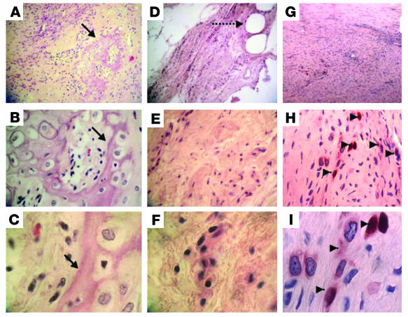
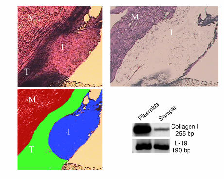
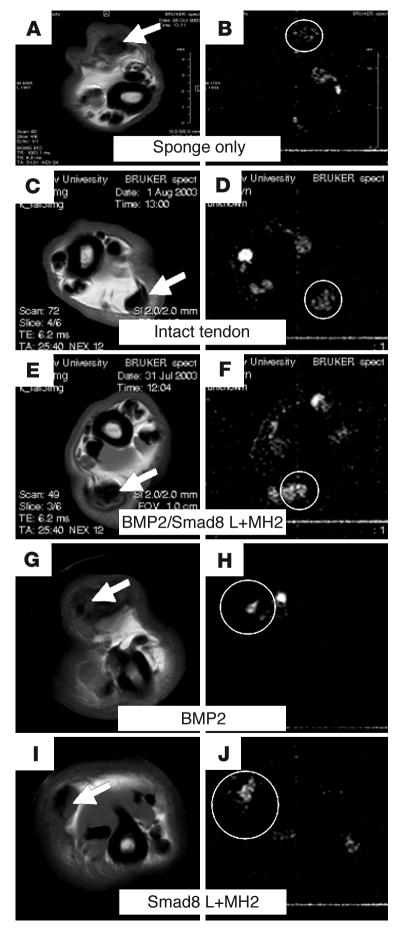
Tissue regeneration requires the recruitment of adult stem cells and their differentiation into mature committed cells. In this study we describe what we believe to be a novel approach for tendon regeneration based on a specific signalling molecule, Smad8, which mediates the differentiation of mesenchymal stem cells (MSCs) into tendon-like cells. A biologically active Smad8 variant was transfected into an MSC line that coexpressed the osteogenic gene bone morphogenetic protein 2 (BMP2). The engineered cells demonstrated the morphological characteristics and gene expression profile of tendon cells both in vitro and in vivo. In addition, following implantation in an Achilles tendon partial defect, the engineered cells were capable of inducing tendon regeneration demonstrated by double quantum filtered MRI. The results indicate what we believe to be a novel mechanism in which Smad8 inhibits the osteogenic pathway in MSCs known to be induced by BMP2 while promoting tendon differentiation. These findings may have considerable importance for the therapeutic replacement of tendons or ligaments and for engineering other tissues in which BMP plays a pivotal developmental role.


Figures

Figure 1
Figure 1. Tenogenic phenotype in C3H10T1/2-BMP2 cells due to forced expression of Smad8 L+MH2.
The parental C3H10T1/2 and C3H10T1/2 constitutively expressing the BMP2 gene were stably transfected with different Smad molecules as described in Methods. (A) Histological analysis of ALP-positive cells at 6 and 10 days after confluence (dac). Note the elongated tenocyte-like appearance of BMP2/Smad8 L+MH2–expressing cells compared with BMP2/Smad8WT–expressing cells. (B) Recombinant expression of FLAG-tagged Smad8 variants in C3H10T1/2-BMP2 cells. Western immunoblotting of cellular extracts of C3H10T1/2-BMP2 cells. Open arrowhead indicates a nonspecific band; filled arrowhead indicates Smad8WT and Smad8 L+MH2 domains. WB, Western blot. (C) Smad8 L+MH2, but not Smad1 or Smad5 L+MH2, elicited a tenocyte-like morphology in C3H10T1/2-BMP2. Histological analysis (ALP) and phase-contrast microscopy. All cells represent day 10 after confluence. Top panels: In the presence of BMP2 all C3H10T1/2 cells underwent mesenchymal differentiation and extensive matrix development resulting in multilayer growth. At this relatively late stage of cultivation residual ALP-positive cells were observed embedded in matrix and on the top of the layer. The presence of coexpressed L+MH2 domains from Smad1 and Smad5 did not influence this morphology. The presence of Smad8 L+MH2 dramatically changed this morphology to an elongated cell phenotype. Bottom panels: Parental C3H10T1/2 cells expressing Smad1 and Smad5 L+MH2 developed an osteoblast-like appearance. Smad8 L+MH2–expressing cells did not possess a comparable osteogenic capacity. Also, development of an elongated cell phenotype was not observed. Magnification, ×40.
Figure 2
Figure 2. Smad8 L+MH2 elicits a tenocyte-like morphology in C3H10T1/2-BMP2 cells, but not in C3H10T1/2-TGFβ1 or C3H10T1/2-GDF5 cells.
Histological analysis (ALP) and phase-contrast microscopy. All cells represent day 11 after confluence. As shown in Figure 1A, the expression of Smad8 L+MH2 led to an elongated tenocyte-like phenotype. This phenotype was not observed when Smad8 L+MH2 was expressed in parental C3H10T1/2 cells or in cells that stably express TGFβ1 or GDF5. TGFβ1 expression led to a dense multilayer growth, but neither osteo- and/or chondrogenic differentiation in vitro nor the formation of tenocyte-like cells in the presence of Smad8 L+MH2 was observed. Stable expression of GDF5 only allowed monolayer growth exhibiting large cellular phenotypes. Smad8 L+MH2 expression did not change this cellular phenotype. RT-PCR analyses show recombinant expression of TGFβ1 or GDF5 in the various cell lines. Western blot analyses show recombinant expression of Smad8 L+MH2 in the recombinant C3H10T1/2 lines.
Figure 4
Figure 4. Smad8 exhibits a low activation potential by various ligands of the TGFβ/BMP family.
(A) FLAG-tagged Smad1 or Smad8 were transiently expressed in C3H10T1/2, and BMP2, TGFβ1, or GDF5 were added for 30 minutes at the indicated concentrations. After Western blotting, ligand-dependent phosphorylation of Smads was shown by antibodies specific for pSmad1, -5, and -8. The expression rate of the Smads was determined by Western blotting using anti-FLAG antibodies. (B) Evaluation of the Smad1 and Smad8 transactivation potential in HEK 293T cells, in which a GAL4 reporter with the GAL4 DNA-binding domain was fused to Smad1WT and Smad8WT proteins as described in Figure 3B. Pooled data from 3 independent experiments are presented. BMP2 led to efficient activation of the GAL4-Smad1 fusion protein, GDF5 was less effective, and TGFβ1 did not exhibit notable activation. The GAL4-Smad8 fusion protein was activated, albeit to a markedly lower extent, by BMP2, but not by TGFβ1 or GDF5. The expression level of the GAL4-Smad fusions was comparable as assessed by Western analyses of cellular extracts and blotting with anti-GAL4 antibodies. (C) Compared with Smad1, Smad8 exhibited a lower BMP2-dependent activation potential in C3H10T1/2. FLAG-tagged Smad1 or Smad8 were transiently expressed in C3H10T1/2, and BMP2 (200 ng/ml) was added for 30 minutes. Top: After Western blotting, BMP2-dependent phosphorylation of Smads is shown by anti-pSmad1, -5, and -8 antibodies. Bottom: Expression rates of the Smads were determined by Western blotting using anti-FLAG antibodies. In the graph at right, the level of BMP2-dependent Smad activation in C3H10T1/2 was evaluated by densitometric scanning.
Figure 3
Figure 3. Smad8 is activated by TGF-β/BMP type I receptors.
(A) Schematic representation of WT Smads and the Smad domains used for functional studies in HEK 293T and C3H10T1/2 cells. MH1 and MH2 are the major conserved Smad domains; “L” shows the linker region between them. The linker region is considerably smaller in Smad8 than in Smad1. (B) Results of reporter assays in HEK 293T cells, in which a GAL4 reporter with the GAL4 DNA-binding domain (GAL4DBD) fused to various forms of Smad proteins was used. Results are expressed as relative luciferase units normalized to β-gal activity and presented as percent of Smad L+MH2, which was arbitrarily set as 100%. Pooled data from at least 3 independent experiments are presented. Smad8 MH2 and L+MH2 domains exhibited a constitutive active transactivating potential comparable to that of Smad1. (C) Activation potential of Smad8 and Smad1 signalling mediators by constitutively active TGF-β/BMP receptors (ALK1–ALK7) in HEK 293T cells. FLAG-tagged Smad8 or Smad1 was transiently coexpressed with constitutively active HA-tagged type I receptors. Expression of all type I receptors was mediated by the identical vector (pcDNA3). Expression rate of the receptors was determined by Western blotting using anti-HA antibodies. Receptor bands indicate variations in the glycosylation of the ectodomain. Type I receptor–dependent phosphorylation of Smads was shown by anti-pSmad1, -5, and -8 antibodies, which also react with phosphorylated Smad8. Smad1 and Smad8 were phosphorylated by most type I receptors. In contrast to Smad1, Smad8 was also efficiently phosphorylated by ALK4 and ALK7 (TGFβ1 receptors).
Figure 5
Figure 5. RT-PCR analyses of the expression levels of chondrogenic, osteogenic, and tenogenic markers in murine C3H10T1/2 cells.
Collagen Ia1, the PTH/PTHrP receptor, and osteocalcin are markers predominantly associated with osteogenesis, and collagen IIa1 is a marker associated with chondrogenic differentiation. Collagen Ia1, six1, six2, scleraxis, eya1, and EphA4 are tenogenic markers. Hypoxanthine guanine phosphoribosyl transferase (HPRT) was used to standardize e PCR conditions.
Figure 6
Figure 6. C3H10T1/2-BMP2/Smad8 L+MH2 cells induce ectopic tendon formation after s.
. implantation. The forced expression of Smad8 L+MH2 in C3H10T1/2-BMP2 cells led to differentiation into tenocytes at murine ectopic sites 30 days after s.c. cell transplantation. (AH) H&E-stained sections of ectopically implanted MSCs. (A and B) C3H10T1/2 progenitors. (C and D) C3H10T1/2-BMP2 cells. (E and F) C3H10T1/2-Smad8 L+MH2 cells. (G and H) C3H10T1/2-BMP2/Smad8 L+MH2 s.c. implants. Magnification, ×40 (top panels), ×100 (bottom panels). C3H10T1/2-BMP2/Smad8 L+MH2 implants demonstrated a completely different morphology than the other implants, highly resembling neotendon tissue. C3H10T1/2 progenitors and C3H10T1/2-Smad8 L+MH2 showed nonspecific mesenchymal tissue morphology, while the C3H10T1/2-BMP2 implants contained cartilage and bone foci. (I) RT-PCR analysis of C3H10T1/2-BMP2/Smad8 L+MH2 cell implants 30 days after implantation. Lane 1, DNA ladder; lanes 2 and 3, engineered cell implants; lane 4, mouse ligament tissue. All 3 genes are associated with tendon/ligament tissues. Results obtained from the other implants showed only Collagen I expression and no expression of Six1 and Elastin genes (not shn).
Figure 7
Figure 7. Achilles tendon regeneration model.
An Achilles tendon partial-thickness defect was created in athymic rats. Adult athymic rats (4 months old) were anesthetized as described in Methods. The Achilles tendon (indicated by arrow) was separated from the plantaris and soleus tendons (A), and a 3-mm partial-resection defect was created in its lateral substance (B, arrows indicate the span of the defect). Cells (3 × 10⁶) were seeded onto a collagen sponge (arrow), which was then placed within the tendon defect and sutured to the tendon (C). The skin was closed in a routine manner using 2-0 Mersilk. (D) Noninvasive monitoring of cell survival in the implantation site. C3H10T1/2-BMP2/Smad8 L+MH2 progenitor cells expressing the luciferase gene were implanted in an Achilles tendon defect in the rat. CCCD analysis demonstrated a positive luciferase signal detected in the implantation site (arrow), indicating the survival of implanted cls.
Figure 8
Figure 8. Neotendon tissue formation in a partial-defect model in the Achilles tendon of a rat.
C3H10T1/2-BMP2 (AC), C3H10T1/2-Smad8 L+MH2 (DF), and C3H10T1/2-BMP2/Smad8 L+MH2 cells infected with adeno-LacZ (GI) were implanted in an Achilles tendon defect in nude rats (3 × 10⁶ cells per implant). (AF) Histological (H&E) and (GI) immunohistochemical (anti-LacZ) staining was performed 4 weeks after implantation. (AC) Tendon implanted with C3H10T1/2-BMP2 cells demonstrated cartilage and bone foci formation within the tendon tissue (arrows). (DF) Tendon implanted with C3H10T1/2-Smad8 L+MH2 cells showed mesenchyme-like tissue formation at the site of implantation (dashed arrow shows suture used to hold collagen scaffold in the defect site). (GI) Tendon implanted with C3H10T1/2-BMP2/Smad8 L+MH2 cells infected with adeno-LacZ prior to implantation showed LacZ-positive cells in the tenogenic implant (arrowheads). Cells seemed to be arranged parallel to the tendon long axis. Magnification, ×10 (top panels), ×40 (middle panels), ×100 (bottom panels).
Figure 9
Figure 9. Collagen I mRNA is expressed in the site of C3H10T1/2-BMP2/Smad8 L+MH2 progenitor cell implantation in the Achilles tendon.
Upper left panel depicts the implantation site in the host Achilles tendon as viewed under a light microscope. Lower left panel shows a schematic representation of the different histological components in the field of view. Upper right panel depicts the histological section after the implantation region was removed from the Achilles tendon using LCM. Blots show the gel electrophoresis results of RT-PCR performed on RNA extracted from the microdissected sample. Ribosomal protein L-19 served as a control. M, muscle; T, host tendon; I, implantation area. Magnification, ×4.
Figure 10
Figure 10. Tendon-defect repair demonstrated by micro-MRI.
(A, C, E, G, and I) MSME axial sections of Achilles tendon of a rat. (B, D, F, H, and J) DQF images of the same axial sections. Tendon was either implanted with collagen sponge without cells (A and B), left intact (C and D), or implanted with C3H10T1/2-BMP2/Smad8 L+MH2 cells (E and F), C3H10T1/2-BMP2 cells (G and H), or C3H10T1/2-Smad8 L+MH2 cells (I and J). DQF images showed ordered collagen fiber formation in the defect site implanted with C3H10T1/2-BMP2/Smad8 L+MH2 cells at a higher level than that found in the contralateral tendon or in the other experimental groups. Arrows indicate the Achilles tendon at the site of implantation. Circles highlight the DQF signal at the site of implantation or, in the case of no treatment, at the tendon.

Comment in

References

    1. Pennisi E. Tending tender tendons. Science. 2002;2955:1011. - PubMed
    1. Jackson D.W., Simon T.M., Kurzweil P.R., Rosen M.A. Survival of cells after intra-articular transplantation of fresh allografts of the patellar and anterior cruciate ligaments. DNA-probe analysis in a goat model. J. Bone Joint Surg. Am. 1992;74:112–118. - PubMed
    1. Sabiston P., Frank C., Lam T., Shrive N. Allograft ligament transplantation. A morphological and biochemical evaluation of a medial collateral ligament complex in a rabbit model. Am. J. Sports Med. 1990;18:160–168. - PubMed
    1. Olson E.J., et al. The biochemical and histological effects of artificial ligament wear particles: in vitro and in vivo studies. Am. J. Sports Med. 1988;16:558–570. - PubMed
    1. Woods G.A., Indelicato P.A., Prevot T.J. The Gore-Tex anterior cruciate ligament prosthesis. Two versus three year results. Am. J. Sports Med. 1991;19:48–55. - PubMed

Publication types

MeSH terms