. 2006 Apr;116(4):953-60.
doi: 10.1172/JCI25091.

In humans, early cortisol biosynthesis provides a mechanism to safeguard female sexual development

Affiliations

In humans, early cortisol biosynthesis provides a mechanism to safeguard female sexual development

Masahiro Goto et al. J Clin Invest. 2006 Apr.

Abstract

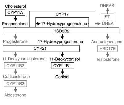
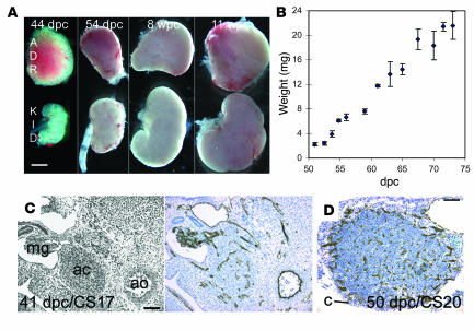
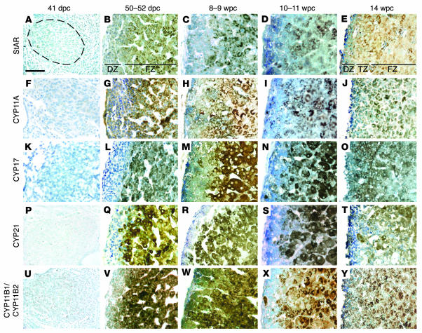
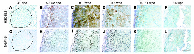
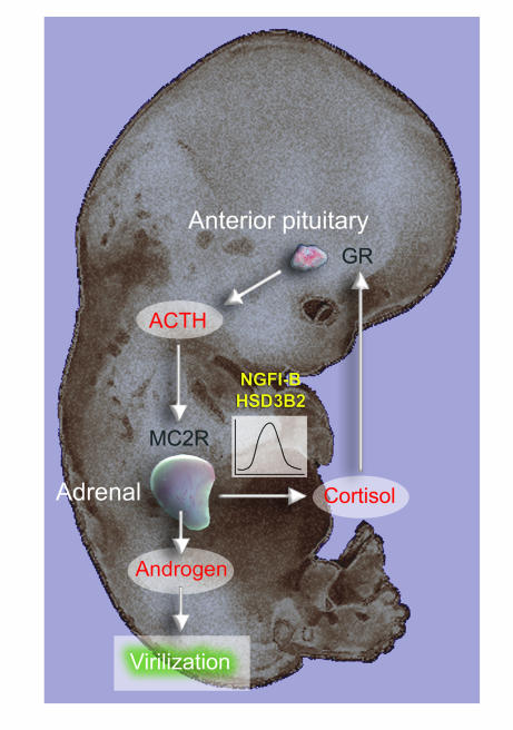
In humans, sexual differentiation of the external genitalia is established at 7-12 weeks post conception (wpc). During this period, maintaining the appropriate intrauterine hormone environment is critical. In contrast to other species, this regulation extends to the human fetal adrenal cortex, as evidenced by the virilization that is associated with various forms of congenital adrenal hyperplasia. The mechanism underlying these clinical findings has remained elusive. Here we show that the human fetal adrenal cortex synthesized cortisol much earlier than previously documented, an effect associated with transient expression of the orphan nuclear receptor nerve growth factor IB-like (NGFI-B) and its regulatory target, the steroidogenic enzyme type 2 3beta-hydroxysteroid dehydrogenase (HSD3B2). This cortisol biosynthesis was maximal at 8-9 wpc under the regulation of ACTH. Negative feedback was apparent at the anterior pituitary corticotrophs. ACTH also stimulated the adrenal gland to secrete androstenedione and testosterone. In concert, these data promote a distinctive mechanism for normal human development whereby cortisol production, determined by transient NGFI-B and HSD3B2 expression, provides feedback at the anterior pituitary to modulate androgen biosynthesis and safeguard normal female sexual differentiation.


Figures

Figure 1
Figure 1. Steroidogenic pathways involving the human adrenal cortex.
De novo cortisol biosynthesis is shown in black; other pathways are in gray. DHEA, dehydroepiandrosterone; DHEAS, DHEA sulfate; ST, sulfotransferase.
Figure 2
Figure 2. Growth and vascular development of the human early fetal adrenal gland.
(A) Relative proportion of adrenal gland (ADR) to kidney (KID) during early development. (B) Weight (α SEM) of individual adrenal glands and developmental age. (C and D) Brightfield IHC with anti-CD34 to demonstrate vascular development counterstained by toluidine blue at 41 (C) and 50 (D) dpc. An adjacent H&E section is shown at the left of C. ac, adrenal cortex; ao, aorta; c, capsule; mg, mesonephric glomerulus. Scale bars: 1 mm (A), 300 μm (C and D).
Figure 3
Figure 3. Steroidogenic enzyme and StAR expression in the developing human adrenal gland.
Brightfield IHC at sequential developmental ages counterstained by toluidine blue with antibodies to StAR (AE), CYP11A (FJ), CYP17 (KO), CYP21 (PT), and CYP11B1/CYP11B2 (UY). Dotted ring in A illustrates extent of the adrenal cortex in left panels (A, F, K, P, and U). In other images, the definitive zone (DZ) is oriented to the left and the fetal zone (FZ) to the right. At 14 wpc, these 2 zones were separated by an additional transitional zone (TZ). Scale bar: 300 μm.
Figure 4
Figure 4. HSD3B and NGFI-B immunoreactivity in the developing human adrenal gland.
Brightfield IHC with antibodies to HSD3B (AF) and NGFI-B (GL) counterstained with toluidine blue. Dotted rings in A and G illustrate extent of the adrenal cortex. In other images, the definitive zone is oriented to the left and fetal zone to the right. Scale bar: 300 μm.
Figure 5
Figure 5. RT-PCR analysis of adrenocortical enzymes and StAR at 8 wpc.
(A) RT followed by 22 and 28 cycles of PCR for transcripts encoding steroidogenic enzymes. (B) Specific identification of HSD3B2 and HSD3B1 isoforms in the presence (+) and absence (–) of RT. No HSD3B1 transcript was detected in the adrenal sample after 42 cycles of PCR. Positive controls were fetal testis (HSD3B2) and skin (HSD3B1); negative controls were H2O and genomic DNA (G).
Figure 6
Figure 6. Cortisol content and secretion from the early human adrenal cortex.
(A) Cortisol content (mean α SEM) per mg adrenal tissue at 8, 9, and 10 wpc, with 8 wpc kidney as control. (B) Cortisol secretion (mean α SEM) of paired adrenal glands stimulated by 10 μM forskolin at 8, 9, and 10 wpc. (C) Cortisol secretion (mean α SEM) of paired adrenal glands at 8 wpc in response to varying doses of ACTH(1–24). Statistical analyses of stimulated secretion compared to basal secretion achieved the same level of significance (***P < 0.005) for each ACTH dose. *P < 0.05; **P < 0.02. (D) PCR amplification of type 2 melanocortin receptor (MC2R) in the presence (+) and absence (–) of RT in the adrenal gland, testis, and ovary at 8 wpc with GAPDH conol.
Figure 7
Figure 7. Human early anterior pituitary development.
(A) Sagittal section from head at 50 dpc. Pound symbol indicates oral cavity. (BD) Brightfield IHC with antibodies to ACTH (B and D) and GR (C) counterstained by toluidine blue. B and C show higher-magnification views of boxed region in A. Arrows show overlapping expression profiles of cytoplasmic ACTH and nuclear GR in adjacent sections. (D) Anterior pituitary at 8 wpc. bs, basosphenoid bone; h, developing hypothalamus; rp, Rathke’s pouch; t, tongue. Scale bars: 500 μm (A), 100 μm (B and C), 200 μm (D).
Figure 8
Figure 8. Virilization of the external genitalia and adrenal androgen biosynthesis.
(A) Undifferentiated human external genitalia at 8 wpc. (B) Male differentiation of scrotal folds and fusion of the urethral folds (asterisks mark patent regions, either side) at 10 wpc. gs, genital swelling; gt, genital tubercle; sf, scrotal folds; uf, urethral folds. Scale bars: 500 μm. (C) PCR (35 cycles) in the presence (+) or absence (–) of RT for HSD17B isoforms known to convert androstenedione to testosterone. HSD17B5 is also known as AKR1C3.
Figure 9
Figure 9. Schematic diagram of human early adrenal function and its implications for androgen-mediated development.

Comment in

Similar articles

Cited by

References

    1. White P.C., Speiser P.W. Congenital adrenal hyperplasia due to 21-hydroxylase deficiency. Endocr. Rev. 2000;21:245–291. - PubMed
    1. David M., Forest M.G. Prenatal treatment of congenital adrenal hyperplasia resulting from 21-hydroxylase deficiency. J. Pediatr. . 1984;105:799–803. - PubMed
    1. Mesiano S., Jaffe R.B. Developmental and functional biology of the primate fetal adrenal cortex. Endocr. Rev. 1997;18:378–403. - PubMed
    1. Pepe G.J., Albrecht E.D. Regulation of the primate fetal adrenal cortex. Endocr. Rev. 1990;11:151–176. - PubMed
    1. Narasaka T., Suzuki T., Moriya T., Sasano H. Temporal and spatial distribution of corticosteroidogenic enzymes immunoreactivity in developing human adrenal. Mol. Cell. Endocrinol. . 2001;174:111–120. - PubMed

Publication types

MeSH terms