. 2006 Apr;116(4):961-73.
doi: 10.1172/JCI25422.

Antibodies against citrullinated proteins enhance tissue injury in experimental autoimmune arthritis

Affiliations

Antibodies against citrullinated proteins enhance tissue injury in experimental autoimmune arthritis

Kristine A Kuhn et al. J Clin Invest. 2006 Apr.

Abstract

Antibodies against citrullinated proteins are specific and predictive markers for rheumatoid arthritis although the pathologic relevance of these antibodies remains unclear. To investigate the significance of these autoantibodies, collagen-induced arthritis (CIA) in mice was used to establish an animal model of antibody reactivity to citrullinated proteins. DBA/1J mice were immunized with bovine type II collagen (CII) at days 0 and 21, and serum was collected every 7 days for analysis. Antibodies against both CII and cyclic citrullinated peptide, one such citrullinated antigen, appeared early after immunization, before joint swelling was observed. Further, these antibodies demonstrated specific binding to citrullinated filaggrin in rat esophagus by indirect immunofluorescence and citrullinated fibrinogen by Western blot. To evaluate the role of immune responses to citrullinated proteins in CIA, mice were tolerized with a citrulline-containing peptide, followed by antigen challenge with CII. Tolerized mice demonstrated significantly reduced disease severity and incidence compared with controls. We also identified novel murine monoclonal antibodies specific to citrullinated fibrinogen that enhanced arthritis when coadministered with a submaximal dose of anti-CII antibodies and bound targets within the inflamed synovium of mice with CIA. These results demonstrate that antibodies against citrullinated proteins are centrally involved in the pathogenesis of autoimmune arthritis.


Figures

Figure 1
Figure 1. Anti-CCP antibodies appear prior to visible arthritis and in parallel with the appearance of anti-collagen antibodies.
(A) Six- to 8-week-old DBA/1J mice were untreated or were immunized at day 0 with CII in CFA or CFA alone and then received booster immunizations 21 days later (black arrows). Sera were collected every 7 days and analyzed for antibody levels. Mice were also observed and scored for development of clinical arthritis. Visible signs of arthritis were measured by adding the number of swollen digits on all 4 paws of each mouse. (B) IgG and (D) isotype-specific antibodies against CII were measured by ELISA as described in Methods. (C) IgG and (E) isotype-specific anti-CCP antibodies were measured by ELISA as described in Methods. IgG3 anti-CCP antibodies were below the limits of detection by ELISA and thus are not shown. The data for each mouse within a group are averaged for that group and represented α SEM. For each group, n = 12. Statistical significance was determined using a 1-way ANOVA. *P < 0.05; **P < 0.01; ***P < 0.001 in CIA mice compared with CFA-immunized and unimmunized mice. (F) Sera from Rag1–/– mice (n = 5) were tested and compared with unimmunized DBA/1J (normal mouse serum (NMS); n = 7 for IgM and n = 23 for IgG). Statistical significance was determined using an unpaired 2-tailed Student’s t test. #P = 0.0004 for IgM anti-CCP antibodies in NMS rsus Rag1–/– mice.
Figure 2
Figure 2. A population of antibodies in CIA is specific for the citrullinated proteins filaggrin and fibrinogen.
(A) Sera from an RA patient; (B) CIA, day 35 after immunization; (C) adjuvant control; or (D) an untreated mouse were applied to tissue sections of rat esophagus. Arrows indicate areas of reactivity on epithelium. (E) Mouse sera from arthritic mice on day 35 after immunization were preabsorbed with citrullinated peptide (LXP) or (F) control peptide (LRP). These were then applied to tissue sections of rat esophagus. Serum antibody binding was detected with FITC-conjugated goat anti-mouse IgG. Magnification, ×40. (G) Western blot of unmodified (un) and citrullinated (cit) fibrinogen probed with RA patient or mouse sera. In second and third lanes, proteins are stained with Coomassie to demonstrate relatively equal transfer for Western blot analysis.
Figure 3
Figure 3. Mice tolerized with a citrullinated peptide are significantly protected from CIA.
(A and B) Six- to eight-week-old male DBA/1J mice were tolerized to OVA (n = 19), CII (n = 16), LXP (n = 16), or LKP (n = 12). After challenging these mice with immunization of CII in CFA, disease severity (A) and incidence (B) were measured. (C) Paws from mice in each tolerance group were analyzed for histologic parameters of arthritis as described in Methods. (D) Anti-CII antibodies were measured by ELISA, and (E) antibodies against citrullinated fibrinogen were determined by Western blot. All data are shown as group averages α SEM. Statistical significance was determined by 1-way ANOVA. *P < 0.05; **P < 0.01 in comparison with the OVA and LKP groups. (F) Sera from tolerized mice were applied to synovial antigen arrays to characterize the autoantibody response. The relative reactivities for the labeled antigens in a pool of sera from 9 mice in each LXP- and LKP-tolerized group are shown, as is the clustering of mice based on the relationships of antibody reactivity betwe the mice.
Figure 4
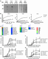
Figure 4. Antibodies specific to citrullinated fibrinogen substantially enhance submaximal arthritis.
(A) Purified D513, 1042, or 1618 monoclonal antibody was applied to a Western blot of unmodified and citrullinated fibrinogen. To compete for antigen binding on the blot, soluble unmodified or citrullinated fibrinogen was added to D513 prior to applying the antibody to the Western blot. (B) The relative reactivities of the monoclonal antibodies for unmodified and citrullinated fibrinogen were determined by applying increasing amounts of biotinylated unmodified and citrullinated fibrinogen (fibr) and CII to ELISA plates coated with monoclonal antibody. The amount of protein binding was detected as described in Methods and is displayed as OD. (C) Additional antigens recognized by D513, 1042, and 1618 were detected using synovial antigen arrays. The relative reactivity for the antigens recognized is shown. (D) Arthrogen, a cocktail of 4 monoclonal antibodies against CII, was transferred intravenously into 6- to 8-week-old male DBA/1J mice at increasing doses followed by 50 μg LPS intraperitoneally 3 days later in order to determine which dose would establish submaximal disease. n = 3 per dose group. (E) D513 (n = 9) or control monoclonal antibody (n = 6) were administered alone or in combination with the submaximal dose of Arthrogen (1 mg). (F and G) Either 1 mg or 2 mg of monoclonal antibody 1042 or 1618 (n = 6) or 2 mg HB5 control monoclonal antibody (n = 6) was combined with 1 mg of Arthrogen and transferred into mice as performed previously with D513. Data shown are the average disease score per group α SEM. Statistical significance was determined using 1-way ANOVA. *P < 0.05; **P < 0.01 in comparison with submaximal Arrogen alone.
Figure 5
Figure 5. D513, 1042, and 1618 monoclonal antibodies bind antigens within the inflamed synovium of mice with CIA.
(A) Immunohistochemistry was performed using 4 μg/ml D513, 1042, and 1618. Antibody binding is indicated by DAB precipitate (brown). Tissue was counterstained with methyl green. A mix of anti-CII monoclonal antibodies and secondary reagents without primary antibody served as negative controls for staining the synovium. Representative serial sections are shown. Magnification, ×100. Arrows point to synovium, and asterisks indicate cartilage. (B) Skin from the site of immunization, draining inguinal LN, and synovium were collected from unimmunized mice, mice immunized with CFA, or mice with CIA at different time points. Western blots of the protein isolated from these tissues were performed with a polyclonal antibody that recognizes the amino acid citrulline or (C) with the monoclonal antibodies 1042, 1618, and D513. Representative blots are shown. Results of Western blots probed with antibodies 1618 and D513 were essentially identical to 1042 and, thus, are not shown.
Figure 6
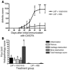
Figure 6. Antibodies against citrullinated fibrinogen can overcome the protective effect of tolerance to citrullinated antigens.
Mice were tolerized with LXP, challenged twice with CII in CFA, and then administered either 2 mg of equal amounts of 1042 and 1618 or 2 mg of the control HB5 antibody on days 22 and 28 after the initial challenge with CII in CFA. (A) Arthritis scores were measured between days 21 and 35, and (B) histologic evaluation was performed on paws collected on day 35. Data are shown as group averages α SEM. Statistical significance was determined by 1-way ANOVA. *P < 0.05; **P < 0.01 in comparison with the LXP plus HB5 control group. Mice tolerized with LKP, challenged with CII in CFA, and given monoclonal antibodies 1042/1618 or HB5 served as additional controls. No difference was detected between these groups and the LXP plus 1042/1618 group (data not sho).

Comment in

References

    1. Arnett F.C., et al. The American Rheumatism Association 1987 revised criteria for the classification of rheumatoid arthritis. Arthritis Rheum. 1988;31:315–324. - PubMed
    1. Aho K., Palusuo T., Kurki P. Marker antibodies of rheumatoid arthritis: diagnostic and pathogenetic implications. Semin. Arthritis Rheum. 1994;23:379–387. - PubMed
    1. Nienhuis R.L., Mandema E. A new serum factor in patients with rheumatoid arthritis; the antiperinuclear factor. Ann. Rheum. Dis. 1964;23:302–305. - PMC - PubMed
    1. Young B.J., Mallya R.K., Leslie R.D., Clark C.J., Hamblin T.J. Anti-keratin antibodies in rheumatoid arthritis. Br. Med. J. 1979;2:97–99. - PMC - PubMed
    1. Vincent C., et al. High diagnostic value in rheumatoid arthritis of antibodies to the stratum corneum of rat oesophagus epithelium, so-called ‘antikeratin antibodies’. Ann. Rheum. Dis. 1989;48:712–722. - PMC - PubMed

Publication types

MeSH terms