

HTLV-1 propels untransformed CD4 lymphocytes into the cell cycle while protecting CD8 cells from death
- PMID: 16585963
- PMCID: PMC1421359
- DOI: 10.1172/JCI27198
HTLV-1 propels untransformed CD4 lymphocytes into the cell cycle while protecting CD8 cells from death
Abstract
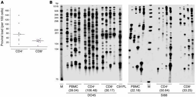
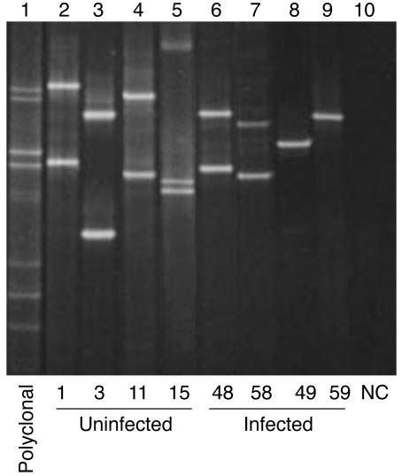
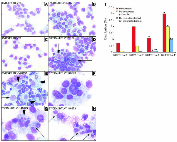
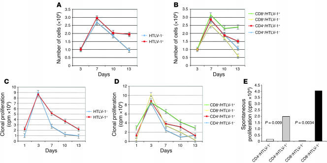
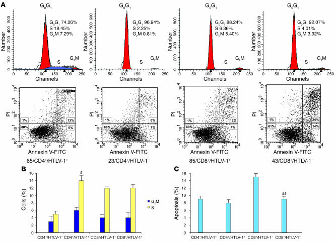
Human T cell leukemia virus type 1 (HTLV-1) infects both CD4+ and CD8+ lymphocytes, yet it induces adult T cell leukemia/lymphoma (ATLL) that is regularly of the CD4+ phenotype. Here we show that in vivo infected CD4+ and CD8+ T cells displayed similar patterns of clonal expansion in carriers without malignancy. Cloned infected cells from individuals without malignancy had a dramatic increase in spontaneous proliferation, which predominated in CD8+ lymphocytes and depended on the amount of tax mRNA. In fact, the clonal expansion of HTLV-1-positive CD8+ and CD4+ lymphocytes relied on 2 distinct mechanisms--infection prevented cell death in the former while recruiting the latter into the cell cycle. Cell cycling, but not apoptosis, depended on the level of viral-encoded tax expression. Infected tax-expressing CD4+ lymphocytes accumulated cellular defects characteristic of genetic instability. Therefore, HTLV-1 infection establishes a preleukemic phenotype that is restricted to CD4+ infected clones.
Figures
Comment in
-
Adult T cell leukemia: a tale of two T cells.J Clin Invest. 2006 Apr;116(4):858-60. doi: 10.1172/JCI28140. J Clin Invest. 2006. PMID: 16585953 Free PMC article.
Similar articles
-
Tax gene expression and cell cycling but not cell death are selected during HTLV-1 infection in vivo.Retrovirology. 2010 Mar 11;7:17. doi: 10.1186/1742-4690-7-17. Retrovirology. 2010. PMID: 20222966 Free PMC article.
-
Persistent clonal proliferation of human T-lymphotropic virus type I-infected cells in vivo.Cancer Res. 1997 Nov 1;57(21):4862-7. Cancer Res. 1997. PMID: 9354450
-
In vivo cellular tropism of human T-lymphotropic virus type II is not restricted to CD8+ cells.Virology. 1995 Jul 10;210(2):441-7. doi: 10.1006/viro.1995.1360. Virology. 1995. PMID: 7542419
-
Clonal expansion of HTLV-1 infected cells depends on the CD4 versus CD8 phenotype.Front Biosci (Landmark Ed). 2009 Jan 1;14(10):3935-41. doi: 10.2741/3502. Front Biosci (Landmark Ed). 2009. PMID: 19273324 Review.
-
HTLV-1 and associated adult T-cell leukemia/lymphoma.Rev Clin Exp Hematol. 2003 Dec;7(4):336-61. Rev Clin Exp Hematol. 2003. PMID: 15129647 Review.
Cited by
-
Tax gene expression and cell cycling but not cell death are selected during HTLV-1 infection in vivo.Retrovirology. 2010 Mar 11;7:17. doi: 10.1186/1742-4690-7-17. Retrovirology. 2010. PMID: 20222966 Free PMC article.
-
A genetic IFN/STAT1/FAS axis determines CD4 T stem cell memory levels and apoptosis in healthy controls and Adult T-cell Leukemia patients.Oncoimmunology. 2018 Feb 13;7(5):e1426423. doi: 10.1080/2162402X.2018.1426423. eCollection 2018. Oncoimmunology. 2018. PMID: 29721391 Free PMC article.
-
Human T-cell leukemia virus type 1 (HTLV-1) and leukemic transformation: viral infectivity, Tax, HBZ and therapy.Oncogene. 2011 Mar 24;30(12):1379-89. doi: 10.1038/onc.2010.537. Epub 2010 Nov 29. Oncogene. 2011. PMID: 21119600 Free PMC article. Review.
-
Time-course of host cell transcription during the HTLV-1 transcriptional burst.PLoS Pathog. 2022 May 16;18(5):e1010387. doi: 10.1371/journal.ppat.1010387. eCollection 2022 May. PLoS Pathog. 2022. PMID: 35576236 Free PMC article.
-
Protein Profile of Blood Monocytes is Altered in HTLV-1 Infected Patients: Implications for HAM/TSP Disease.Sci Rep. 2018 Sep 25;8(1):14354. doi: 10.1038/s41598-018-32324-2. Sci Rep. 2018. PMID: 30254298 Free PMC article.
References
-
- Moules V., et al. Fate of premalignant clones during the asymptomatic phase preceding lymphoid malignancy. Cancer Res. 2005;65:1234–1243. - PubMed
-
- Yoshida M. Multiple viral strategies of Htlv-1 for dysregulation of cell growth control. Annu. Rev. Immunol. 2001;19:475–496. - PubMed
-
- Poiesz B.J., Ruscetti F.W., Reitz M.S., Kalyanaraman V.S., Gallo R.C. Isolation of a new type C retrovirus (HTLV) in primary uncultured cells of a patient with Sezary T-cell leukemia. Nature. 1981;294:268–271. - PubMed
-
- Kalyanaraman V.S., et al. A new subtype of human T-cell leukemia virus (HTLV-II) associated with a T-cell variant of hairy cell leukemia. Science. 1982;218:571–573. - PubMed
Publication types
MeSH terms
LinkOut - more resources
Full Text Sources
Research Materials
