. 2006 Apr;116(4):984-95.
doi: 10.1172/JCI25790.

Nrf2 is a critical regulator of the innate immune response and survival during experimental sepsis

Affiliations

Nrf2 is a critical regulator of the innate immune response and survival during experimental sepsis

Rajesh K Thimmulappa et al. J Clin Invest. 2006 Apr.

Abstract

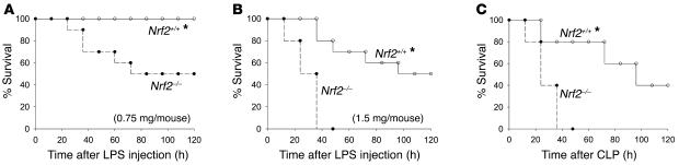
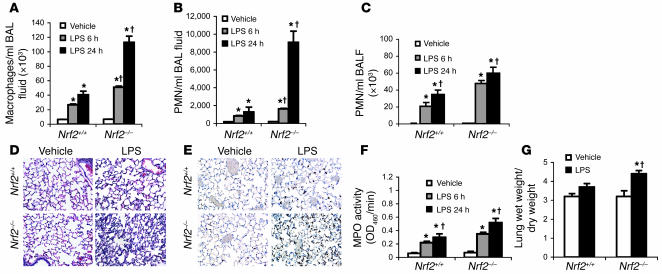
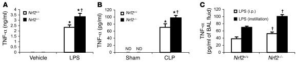
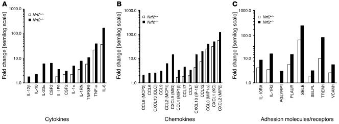
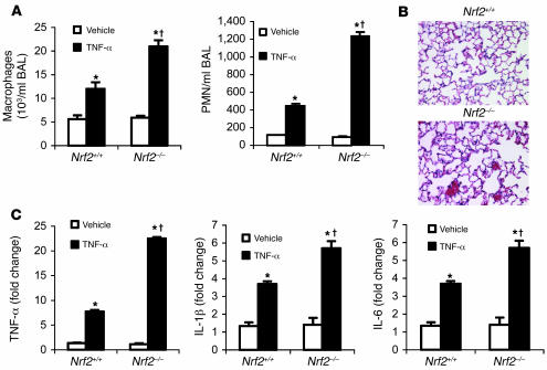
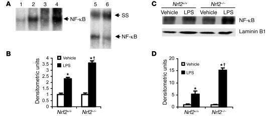
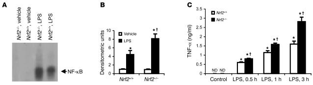
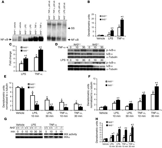
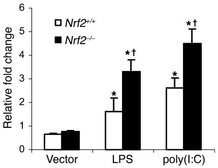
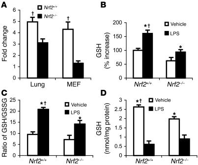
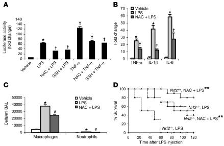
Host genetic factors that regulate innate immunity determine susceptibility to sepsis. Disruption of nuclear factor-erythroid 2-related factor 2 (Nrf2), a basic leucine zipper transcription factor that regulates redox balance and stress response, dramatically increased the mortality of mice in response to endotoxin- and cecal ligation and puncture-induced septic shock. LPS as well as TNF-alpha stimulus resulted in greater lung inflammation in Nrf2-deficient mice. Temporal analysis of pulmonary global gene expression after LPS challenge revealed augmented expression of large numbers of proinflammatory genes associated with the innate immune response at as early as 30 minutes in lungs of Nrf2-deficient mice, indicating severe immune dysregulation. The expression profile indicated that Nrf2 has a global influence on both MyD88-dependent and -independent signaling. Nrf2-deficient mouse embryonic fibroblasts showed greater activation of NF-kappaB and interferon regulatory factor 3 in response to LPS and polyinosinic-polycytidylic acid [poly(I:C)] stimulus, corroborating the effect of Nrf2 on MyD88-dependent and -independent signaling. Nrf2's regulation of cellular glutathione and other antioxidants is critical for optimal NF-kappaB activation in response to LPS and TNF-alpha. Our study reveals Nrf2 as a novel modifier gene of sepsis that determines survival by mounting an appropriate innate immune response.


Figures

Figure 1
Figure 1. Nrf2–/– mice were more sensitive to LPS and septic peritonitis–induced septic shock.
(A and B) Mortality after LPS administration. Age-matched male Nrf2+/+ (n = 10) and Nrf2–/– mice (n = 10) were injected i.p. with LPS (0.75 and 1.5 mg per mouse). (C) Acute septic peritonitis was induced by CLP. CLP and sham operations were performed as described in Methods on age-matched male Nrf2+/+ (n = 10) and Nrf2–/– mice (n = 10). Mortality was assessed every 12 hours for 5 days. *Nrf2+/+ mice showed improved survival compared wi Nrf2–/– mice. P < 0.05.
Figure 2
Figure 2. Nonlethal dose of LPS induced greater lung inflammation in Nrf2-deficient lungs.
(A and B) BAL fluid analysis of Nrf2–/– and Nrf2+/+ mice after 6 and 24 hours of i.p. injection of LPS (60 μg per mouse). (C) BAL fluid analysis of Nrf2–/– and Nrf2+/+ mice after 6 hours and 24 hours of LPS instillation (10 μg per mouse). (D) Histopathological analysis of lungs by H&E staining 24 hours after instillation of LPS. Arrows indicate accumulation of inflammatory cells in the alveolar spaces. Magnification, ×20. (E) Immunohistology of lungs of both genotypes using anti-mouse neutrophil antibody 24 hours after LPS instillation. Sections were counterstained with hematoxylin. Arrows indicate neutrophils. Magnification, ×40. (F) Myeloperoxidase (MPO) activity in lung homogenates of both genotypes 6 and 24 hours after LPS instillation. (G) Pulmonary edema was assessed by the ratio of wet to dry lung weight 24 hours after LPS instillation. Data are presented as mean α SEM; n = 5. *Differs from vehicle control of the same genotype. Differs from LPS-treated wild-type mic P < 0.05.
Figure 3
Figure 3. LPS and CLP induce greater secretion of TNF-α in Nrf2-deficient mice.
(A) Serum concentration of TNF-α in Nrf2+/+ and Nrf2–/– mice 1.5 hours after LPS injection (1.5 mg per mouse). (B) Serum concentration of TNF-α in Nrf2+/+ and Nrf2–/– mice 6 hours after CLP. (C) TNF-α levels in BAL fluid at 2 hours after LPS delivery by i.p. injection (60 μg per mouse) and/or intratracheal instillation (10 μg per mouse). TNF-α in the BAL fluid of vehicle-treated mice was not detectable. Data are presented as mean α SEM. *Differs from vehicle control of the same genotype. Differs from LPS-treated wild-type mice. P < 0.05. ND, n detected.
Figure 4
Figure 4. Greater expression of proinflammatory genes associated with the innate immune response in the lungs of Nrf2-deficient mice.
(AC) Expression of cytokines (A), chemokines (B), and adhesion molecules/receptors (C) 30 minutes after nonlethal i.p. injection of LPS (60 μg per mouse) in Nrf2-deficient and wild-type mice. Gene expression data were obtained from microarray analysis. Data are represented as mean fold change obtained from comparing LPS-challenged to vehicle-treated lungs of the same genotype on a semilog scale. All the represented fold change values of LPS-treated lungs of Nrf2–/– mice are significant compared with wild-type mice at P <0.05.
Figure 5
Figure 5. TNF-α stimulus induced greater lung inflammation in Nrf2-deficient mice.
(A) BAL fluid analysis at 6 hours after i.p. injection of TNF-α (10 μg/mouse). (B) Histopathological analysis of lungs of Nrf2+/+ and Nrf2–/– mice by H&E staining 24 hours after i.p. injection of TNF-α (10 μg/mouse). Vehicle-treated lungs are not shown. Magnification, ×20. (C) Gene expression analysis of TNF-α, IL-1β, and IL-6 by real-time PCR in the lungs of Nrf2–/– and Nrf2+/+ mice 30 minutes after TNF-α challenge. Data are presented as mean α SEM. *Differs from vehicle control of the same genotype. Differs from LPS-treated wild-type mice. < 0.05.
Figure 6
Figure 6. LPS induced greater NF-κB activation in lungs of Nrf2-deficient mice.
(A) Lung nuclear extracts from Nrf2–/– and Nrf2+/+ mice were assayed for NF-κB DNA-binding by EMSA 30 minutes after instillation of LPS (10 μg per mouse). The major NF-κB bands contained p65 and p55 subunits, as determined by the supershift obtained by p65 and p50 antibody. Lanes: 1, vehicle, Nrf2+/+; 2, LPS, Nrf2+/+; 3, vehicle, Nrf2–/–; 4, LPS, Nrf2–/–; 5, LPS, Nrf2+/+ with p65 antibody, 6, LPS, Nrf2+/+ with p50 antibody. SS, supershift. (B) Quantification of NF-κB DNA-binding was performed by densitometric analysis. All values are mean α SEM obtained from 3 animals per treatment group and are represented as relative to respective vehicle control. (C) Nuclear accumulation of p65 by Western blot in the nuclear extracts derived from lungs of Nrf2+/+ and Nrf2–/– mice 30 minutes after instillation of LPS (10 μg/mouse). Lamin B1 was used as loading control. (D) Densitometric analysis of Western blot of RelA relative to wild-type vehicle control. All values are mean α SEM; n = 3. *Differs from vehicle control of the same genotype. Differs from LPS-treat wild-type mice. P < 0.05.
Figure 7
Figure 7. Lack of Nrf2 augments NF-κB activation in macrophages.
(A) Nuclear extracts of Nrf2+/+ and Nrf2–/– peritoneal macrophages were assayed for NF-κB DNA-binding by EMSA 20 minutes after LPS treatment (1 ng/ml). (B) Densitometric analysis of NF-κB DNA-binding relative to wild-type vehicle control. Values are mean α SEM; n = 3. (C) TNF-α levels in the culture media from Nrf2+/+and Nrf2–/–peritoneal macrophages after 0.5 hours, 1 hours, and 3 hours of LPS treatment (1 ng/ml). *Differs from vehicle control of the same genotype. Differs from wild-type treatment grou P < 0.05.
Figure 8
Figure 8. LPS and/or TNF-α stimulus induces greater NF-κB activation in Nrf2-deficient MEFs.
(A) Nuclear extracts from Nrf2+/+ and Nrf2–/– MEFs were assayed for NF-κB DNA-binding activity by EMSA 30 minutes after LPS (0.5 μg/ml) and or TNF-α (10 ng/ml). The major NF-κB bands contained p65 and p55 subunits, as determined by the supershift analysis using p65 and p55 antibody. (B) Quantification of NF-κB DNA-binding was performed by densitometric analysis. All values are mean α SEM (n = 3) and are represented relative to respective vehicle control. (C) NF-κB–mediated reporter activity in MEFs of both genotypes challenged with LPS (0.5 μg/ml) and TNF-α (10 ng/ml). At 24 hours after transfection with p–NF-κB–Luc vector, cells were treated with LPS and/or TNF-α for 3 hours, and then luciferase activity was measured. Data are mean α SEM from 3 independent experiments (n = 3) and are represented relative to respective vehicle control. (D) Immunoblot of IκB-α and p–IκB-α protein in Nrf2+/+ and Nrf2–/– MEFs after LPS (0.5 μg/ml) or TNF-α (10 ng/ml) stimulus. (E and F) Quantification of IκB-α (E) and p–IκB-α (F) protein in Nrf2+/+ and Nrf2–/– MEFs by densitometric analysis. Data are mean α SEM (n = 3) and are shown as relative to respective vehicle control. (G) IKK activity in Nrf2+/+ and Nrf2–/– MEFs after LPS (0.5 μg/ml) or TNF-α (10 ng/ml) stimulus. (H) Quantification of IKK activity in Nrf2+/+ and Nrf2–/– MEFs by densitometric analysis. Densitometric units are normalized to IKKα. Data are mean α SEM (n = 3) and are relative to respective controls. *Differs from vehicle control of the same genotype. Differs frowild-type treatment group. P < 0.05.
Figure 9
Figure 9. Nrf2 deficiency increases LPS- and or poly(I:C)-induced IRF3-mediated luciferase reporter activity in MEFs.
At 24 hours after transfection with ISRE-Tk-Luc vector, cells were treated with LPS and or poly(I:C) for 6 hours, and luciferase assays were performed 6 hours after treatment. For poly(I:C) stimulation, MEFs were transfected with 6 μg of poly(I:C) in 8 μl of Lipofectamine2000. Data are mean α SEM from 3 independent experiments; n = 3. *Differs from vehicle control of the same genotype; Differs from wild-type treatment group. P <0.05.
Figure 10
Figure 10. Lower levels of GSH in the lungs and MEFs of Nrf2-deficient mice.
(A) Constitutive expression of GCLC in lungs and MEFs of Nrf2+/+ and Nrf2–/– mice. (B) GSH levels in the lungs of mice of both genotypes 24 hours after LPS instillation (10 μg per mouse). Data are mean α SEM from 3 independent experiments and are expressed as percentage increases relative to vehicle-treated Nrf2+/+ group. (C) Ratio of GSH to GSSG measured 24 hours after LPS instillation in the lung of Nrf2+/+ and Nrf2–/– mice. Data are mean α SEM from 3 independent experiments. (D) GSH levels in Nrf2+/+ and Nrf2–/– MEFs at 1 hour after LPS (0.5 μg/ml) or TNF-α (10 ng/ml) stimulus. Data are presented as mean α SEM; n = 4. *Differs from vehicle control of the same genotype. Differs from wild-type treatmt group. P < 0.05.
Figure 11
Figure 11. Pretreatment with exogenous antioxidants alleviates inflammation in Nrf2-deficient mice.
(A) NF-κB–mediated luciferase reporter activity in Nrf2–/– MEFs pretreated for 1 hour with NAC (10 mM) and/or GSH-monoethyl ester (GSH) (1 mM) after 3 hours of LPS (0.5 μg/ml) and/or TNF-α (10 ng/ml) stimulus. Data are presented as mean α SEM (n = 4). *Differs from vehicle control. Differs from group that was treated with LPS or TNF-α only. P < 0.05. (B) Expression of TNF-α, IL-1β, and IL-6 by real-time PCR at 30 minutes in the lungs of Nrf2–/– mice pretreated with NAC after LPS (i.p., 60 μg per mouse) challenge. (C) BAL fluid analysis at 6 hours in lungs of Nrf2–/– mice pretreated with NAC after LPS (i.p., 60 μg per mouse) challenge. Nrf2–/– mice were pretreated with 3 doses of NAC (500 mg/kg body weight, i.p., every 4 hours). Data are presented as mean α SEM. n = 4. #Differs from only LPS treatment. P < 0.05. (D) LPS-induced mortality in Nrf2–/– and Nrf2+/+mice pretreated with NAC. Age-matched male Nrf2–/–(n = 10) and Nrf2+/+ mice (n = 10) were pretreated with NAC (i.p., 500 mg/kg body weight) and/or saline every day for 4 days followed by LPS challenge (1.5 mg per mouse). Mortality was assessed every 12 hours for 5 days. **Mice pretreated with NAC had improved survival comped with vehicle-pretreated mice. P < 0.05.

Comment in

References

    1. Cohen J. The immunopathogenesis of sepsis. Nature. 2002;420:885–891. - PubMed
    1. Pinsky M.R. Dysregulation of the immune response in severe sepsis. Am. J. Med. Sci. 2004;328:220–229. - PubMed
    1. Angus D.C., et al. Epidemiology of severe sepsis in the United States: analysis of incidence, outcome, and associated costs of care. Crit. Care Med. 2001;29:1303–1310. - PubMed
    1. Arcaroli J., Fessler M.B., Abraham E. Genetic polymorphisms and sepsis. Shock. 2005;24:300–312. - PubMed
    1. Thimmulappa R.K., et al. Identification of Nrf2-regulated genes induced by the chemopreventive agent sulforaphane by oligonucleotide microarray. Cancer Res. 2002;62:5196–5203. - PubMed

Publication types

MeSH terms