. 2006 May 1;193(9):1244-9.
doi: 10.1086/502975. Epub 2006 Mar 27.

Anti-severe acute respiratory syndrome coronavirus immune responses: the role played by V gamma 9V delta 2 T cells

Affiliations

Anti-severe acute respiratory syndrome coronavirus immune responses: the role played by V gamma 9V delta 2 T cells

Fabrizio Poccia et al. J Infect Dis. .

Erratum in

  • J Infect Dis. 2006 Jul 15;194(2):267

Abstract

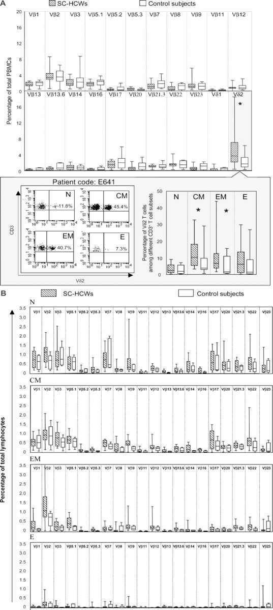
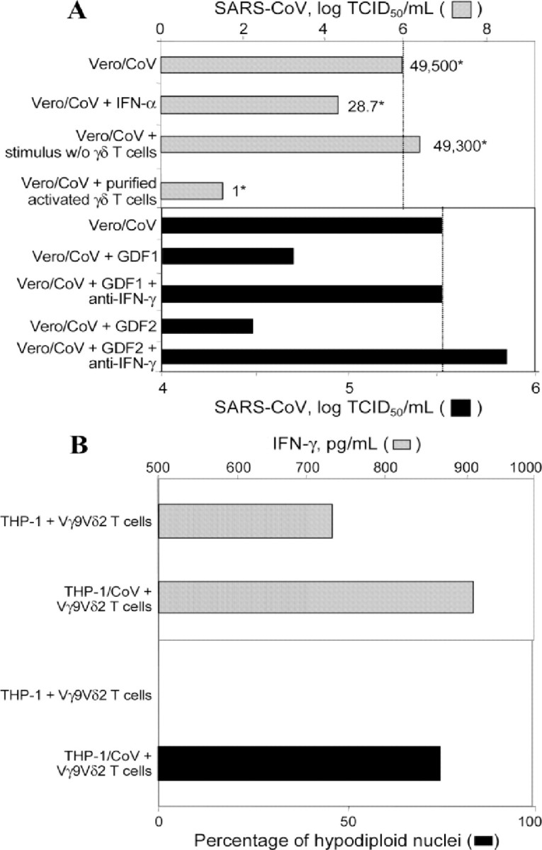
Severe acute respiratory syndrome (SARS) is caused by a novel coronavirus (SARS-CoV) strain. Analyses of T cell repertoires in health care workers who survived SARS-CoV infection during the 2003 outbreak revealed that their effector memory V gamma 9V delta 2 T cell populations were selectively expanded ~3 months after the onset of disease. No such expansion of their alpha beta T cell pools was detected. The expansion of the V gamma 9V delta 2 T cell population was associated with higher anti-SARS-CoV immunoglobulin G titers. In addition, in vitro experiments demonstrated that stimulated V gamma 9V delta 2 T cells display an interferon- gamma -dependent anti-SARS-CoV activity and are able to directly kill SARS-CoV-infected target cells. These findings are compatible with the possibility that V gamma 9V delta 2 T cells play a protective role during SARS.


Figures

Figure 1
Figure 1
Induction of distinct changes in T cell subsets by severe acute respiratory syndrome (SARS) coronavirus (SARS-CoV) infection. A Vβ and Vδ T cell repertoires in health care workers (HCWs) who had had SARS and had convalesced (SC-HCWs; n=15; hatched bars) and in healthy control subjects (n=11; white bars), as assessed by flow cytometry with 22 T cell receptor chain–specific monoclonal antibodies. A representative experiment using peripheral-blood mononuclear cells (PBMCs) from one of the SC-HCWs is shown in the inset, which illustrates the frequencies of Vδ2 cells among naive (N), central memory (CM), effector memory (EM), and effector (E) CD3+ T cells. B Vβ T cell repertoire among the different effector memory αβ T cell subsets. In both panel A and panel B, the bars indicate the interquartile ranges of the individual measurements, the horizontal lines within the boxes indicate the median values, and the vertical lines indicate the ranges of the lowest and highest measurements. Statistical analysis was performed using the nonparametric Mann-Whitney U test. *Statistically significant (P<.05)
Figure 2
Figure 2
Noncytolytic antiviral activity and cytokine production. A Inhibition of severe acute respiratory syndrome (SARS) coronavirus (SARS-CoV) replication by interferon (IFN)–γ–like factors produced by activated Vγ9Vδ2 T cells. The gray and black bars show the log TCID50 per milliliter. One representative experiment (of 3 independent experiments) is shown. The relative numbers of SARS-CoV RNA copies (compared with those in control cultures) are indicated by asterisks and reflect the average of measurements in 3 separate wells. GDF, γδ factor released by Vδ2 T cell lines during the 24-h isopentenyl pyrophosphate–stimulation period; Vero/CoV, Vero cells infected with SARS-CoV. B Ability of SARS-CoV–infected THP-1 cells (THP-1/CoV) to induce IFN-γ production by Vγ9Vδ2 T cells, as determined by ELISA. The amount of IFN-γ in supernatants from 48-h cultures (gray bars) was analyzed in the presence and the absence of Vγ9Vδ2 T cell lines. The spontaneous (background) release of IFN-γ was <7% of the stimulation-induced release and was subtracted from the values measured in the experimental cocultures. In the same experiments, the percentages of hypodiploid target cells were calculated using propidium iodide labeling (black bars) after 24 h of coculture. The relative frequencies of dead THP-1 cells in control cultures without Vγ9Vδ2 T cells never exceeded 15% and were subtracted from the values measured in the experimental cocultures

References

    1. Ksiazek TG, Erdman D, Goldsmith CS, et al. A novel coronavirus associated with severe acute respiratory syndrome. N Engl J Med. 2003;348:1953–66. - PubMed
    1. Lee N, Hui D, Wu A, et al. A major outbreak of severe acute respiratory syndrome in Hong Kong. N Engl J Med. 2003;348:1986–94. - PubMed
    1. Dandekar AA, Perlman S. Virus-induced demyelination in nude mice is mediated by gamma delta T cells. Am J Pathol. 2002;161:1255–63. - PMC - PubMed
    1. Dandekar AA, O’Malley K, Perlman S. Important roles for gamma interferon and NKG2D in gammadelta T-cell-induced demyelination in T-cell receptor beta-deficient mice infected with a coronavirus. J Virol. 2005;79:9388–96. - PMC - PubMed
    1. Gioia C, Horejsh D, Agrati C, et al. T-cell response profiling to biological threat agents including the SARS coronavirus. Int J Immunopathol Pharmacol. 2005;18:525–30. - PubMed

Publication types

MeSH terms

Substances