

Destructive granuloma derived from a liver cyst: a case report
- PMID: 16586558
- PMCID: PMC4124364
- DOI: 10.3748/wjg.v12.i11.1798
Destructive granuloma derived from a liver cyst: a case report
Abstract
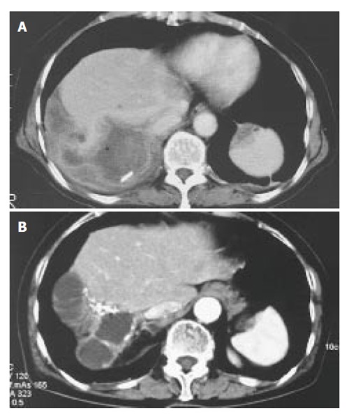
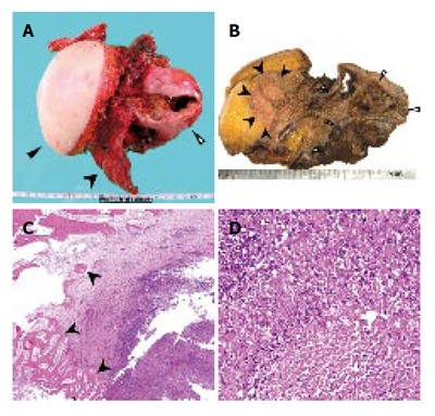
We herein report the case of an idiopathic liver cystic mass which aggressively infiltrated the thoraco-abdominal wall. A 74-year-old woman who had a huge cystic lesion in her right hepatic lobe was transferred to our hospital for further examinations. Imaging studies revealed a simple liver cyst, and the cytological findings of intracystic fluid were negative. She was followed up periodically by computed tomography (CT) scans. Seven years later, she complained of a prominence and dull pain in her right thoraco-abdominal region. CT revealed an enlargement of the cystic lesion and infiltration into the intercostal subcutaneous tissue. We suspected the development of a malignancy inside the liver cyst such as cystadenocarcinoma, and she therefore underwent surgery. A tumor extirpation was performed, including the chest wall, from the 7th to the 10th rib, as well as a right hepatic lobectomy. Pathologically, the lesion consisted of severe inflammatory change with epithelioid cell granuloma and bone destruction without any malignant neoplasm. No specific pathogens were evident based on further histological and molecular examinations. Therefore the lesion was diagnosed to be a destructive granuloma associated with a long-standing hepatic cyst. Since undergoing surgery, the patient has been doing well without any signs of recurrence.
Figures


Similar articles
-
Intracystic hemorrhage of a simple liver cyst mimicking a biliary cystadenocarcinoma.J Gastroenterol. 2003;38(2):190-3. doi: 10.1007/s005350300032. J Gastroenterol. 2003. PMID: 12640536
-
Cystic lesions in the liver: benign or malignant?Acta Chir Belg. 2007 Sep-Oct;107(5):554-6. doi: 10.1080/00015458.2007.11680122. Acta Chir Belg. 2007. PMID: 18074919
-
Cystadenocarcinoma of the liver without mesenchymal stroma: possible progression from a benign cystic lesion suspected by follow-up imagings.J Gastroenterol. 2003;38(6):588-92. J Gastroenterol. 2003. PMID: 12856676
-
[Intracystic hemorrhage of simple hepatic cyst simulating cystadenocarcinoma of the liver--report of two cases].Nihon Shokakibyo Gakkai Zasshi. 1994 Dec;91(12):2264-8. Nihon Shokakibyo Gakkai Zasshi. 1994. PMID: 7837695 Review. Japanese. No abstract available.
-
Squamous cell carcinoma of the liver originating from a solitary non-parasitic cyst case report and review of the literature.HPB Surg. 1996;10(1):45-9. doi: 10.1155/1996/97680. HPB Surg. 1996. PMID: 9187552 Free PMC article. Review.
Cited by
-
Cystic tumors of the liver: a practical approach.World J Gastroenterol. 2008 Jun 21;14(23):3616-20. doi: 10.3748/wjg.14.3616. World J Gastroenterol. 2008. PMID: 18595127 Free PMC article.
-
Hepatic Xanthogranuloma that Originated from a Liver Cyst and Mimicked a Malignant Tumor.In Vivo. 2020 Jul-Aug;34(4):2067-2071. doi: 10.21873/invivo.12009. In Vivo. 2020. PMID: 32606184 Free PMC article.
References
Publication types
MeSH terms
LinkOut - more resources
Full Text Sources
Medical
