. 2006 May;4(5):e112.
doi: 10.1371/journal.pbio.0040112. Epub 2006 Apr 11.

Structure-guided recombination creates an artificial family of cytochromes P450

Affiliations

Structure-guided recombination creates an artificial family of cytochromes P450

Christopher R Otey et al. PLoS Biol. 2006 May.

Abstract

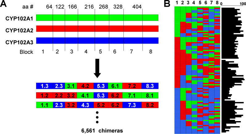
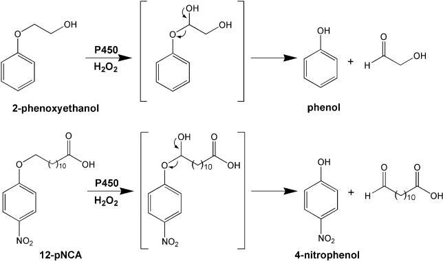
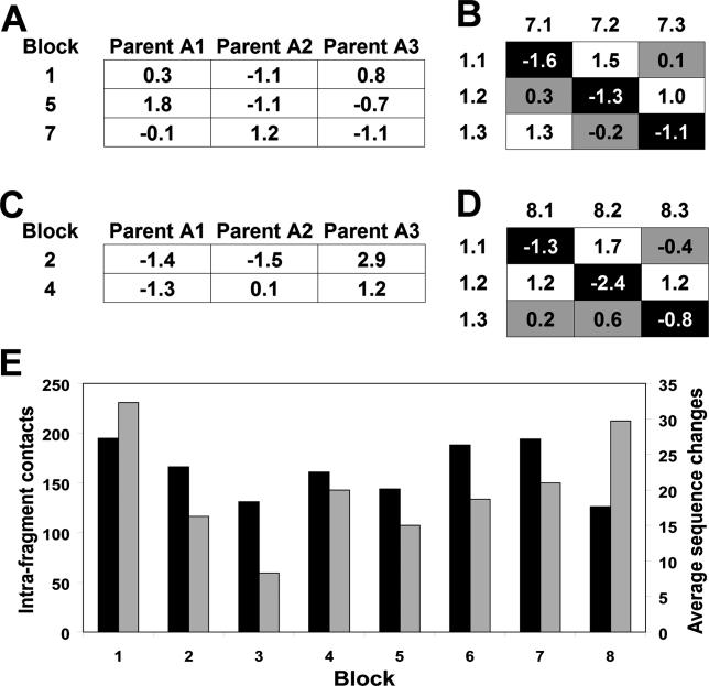
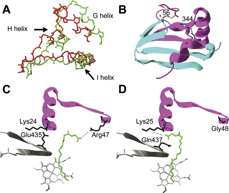
Creating artificial protein families affords new opportunities to explore the determinants of structure and biological function free from many of the constraints of natural selection. We have created an artificial family comprising 3,000 P450 heme proteins that correctly fold and incorporate a heme cofactor by recombining three cytochromes P450 at seven crossover locations chosen to minimize structural disruption. Members of this protein family differ from any known sequence at an average of 72 and by as many as 109 amino acids. Most (>73%) of the properly folded chimeric P450 heme proteins are catalytically active peroxygenases; some are more thermostable than the parent proteins. A multiple sequence alignment of 955 chimeras, including both folded and not, is a valuable resource for sequence-structure-function studies. Logistic regression analysis of the multiple sequence alignment identifies key structural contributions to cytochrome P450 heme incorporation and peroxygenase activity and suggests possible structural differences between parents CYP102A1 and CYP102A2.


Figures

Figure 1
Figure 1. Diverse Chimeras Created by Site-Directed Recombination
(A) Site-directed recombination of three bacterial cytochromes P450 showing crossover sites chosen to minimize the number of disrupted contacts (number is last residue of the sequence block according to CYP102A1 numbering). Blocks are assigned numbers 1 through 8 and three fragments are possible at each block. Three example chimeras are shown to illustrate the fragment nomenclature, e.g., fragment 1.3 is block 1 inherited from parent A3. (B) Sequences of three parents and 97 folded P450 chimeras and number of amino acid changes relative to the closest parent (bar on right).
Figure 2
Figure 2. Structural Model of Heme-Domain Backbone Structure Showing Positions of Each Block
Model is based on the crystal structure of CYP102A1 (2HPD) [ 26]. Blocks are color-coded as shown and heme is shown in CPK coloring.
Figure 3
Figure 3. Comparison of Library Design to Domains, Dynamics, and Secondary Structure of CYP102A1
(A) Crossovers in the library designed using the SCHEMA energy function capture domain boundaries of CYP102A1 determined from molecular dynamics simulations [ 27]. Crossovers between blocks 2–3, 4–5, 5–6, and 7–8 lie within α-helices. (Secondary structure assignment is based on the CYP102A1 crystal structure [ 24]). (B) Plot of the RMSD between the backbone atoms of the substrate-bound (closed) and unbound (open) structures of CYP102A1. The RMSD was calculated by comparing molecule B of the substrate-free structure [ 29] and molecule A of the structure bound to palmitoleic acid [ 26] using Swiss PDB Viewer. Vertical lines designate crossover locations and blocks are numbered. Crossovers between blocks 1–2, 5–6, 6–7, and 7–8 occur at positions that move < 1.2 Å between the two structures. Crossover 3–4 is located next to a region of high identity and may be shifted towards the N-terminus by up to 14 residues and still produce the same chimeras. This shift allows it to occur at a position which moves < 1.2 Å.
Figure 4
Figure 4. Ternary Diagrams Showing the Distribution of Chimera Amino Acid Compositions
(A) Compositions of 955 folded (closed circles) and not-folded (open circles) chimeric sequences. Each data point represents the relative amino acid identity between a chimera and each parental sequence not including positions conserved between all three parents. This distance was calculated by determining the number of amino acids a chimera shares with each parent and dividing by their sum. The three relative identities add up to one. Since each parent shares some sequence identity with the other two, they do not lie at the corners of the diagram. (B) Compositions of 441 chimeras tested for activity on 12-pNCA: active chimeras (closed circles) and not active (open circles). Chimeras composed mostly of A3 and chimeras near the center tend to be inactive on 12-pNCA.
Figure 5
Figure 5. Substrates and Major Products of P450 Peroxygenase Reactions with 2-Phenoxyethanol and p-Nitrophenoxydodecanoic Acid (12-pNCA)
In both cases, hydroxylation yields a hemiacetal which decomposes to phenolic products detectable in high-throughput assays.
Figure 6
Figure 6. LRA of MSAs Identified Blocks and Block Pairs That Contribute to Whether a Chimera Folds and Binds Heme and Whether It Exhibits Activity on 12-pNCA
(A) Intra-fragment terms in the energy model from LRA of folded/not-folded sequences indicate that blocks 1, 5, and 7 make significant contributions to folding and incorporation of heme. Negative energies increase the likelihood of folding and correctly binding heme while positive ones decrease it. (B) The single significant inter-fragment interaction from LRA of folded/not-folded sequences comes from pair 1–7 and includes the nine energy terms for pair 1–7, which can be divided into three groups. The on-diagonal elements (filled black) are the most stabilizing. The three terms filled gray have roughly average energy. The three white elements are destabilizing relative to the others. (C) Significant intra-fragment terms from LRA of the MSA of active/not-active sequences indicate that blocks 2 and 4 have significant effects on peroxygenase activity. (D) The single significant inter-fragment interaction between blocks 1 and 8, showing the nine terms, divided into similar groups as in part B. (E) Black bars, intra-fragment contacts within each block, as defined by the SCHEMA distance of 4.5 Å [ 16]. Gray bars, the average number of sequence changes between each parent.
Figure 7
Figure 7. Structural Elements That Contribute Significantly to Proper Folding and Incorporation of Heme and Model of Substrate Binding in CYP102A1 and CYP102A2
(A) Movement of block 5 between open (red) and closed (green) structural forms based on alignment of heme cofactor. The average displacement over the whole block is 3.6 Å. (B) Residues that could contribute to positively and negatively interacting fragments at blocks 1 and 7. Residue 56 (shown as arginine) is an arginine, glutamate, and glutamine; and residue 344 (shown as glutamate) is a glutamate, lysine, and glutamate in A1, A2, and A3, respectively. The fragment pairs that result in unfavorable charge–charge interactions for these closely spaced side chains are unfavorable overall for folding and heme incorporation. (C) In CYP102A1 the carboxylate group of the fatty acid substrate (in green) interacts with arginine 47 from block 1 (dashed line). Residue 435, from block 8, and residue 24 may form a salt bridge. Portions of blocks 1 and 8 are shown in purple and grey, respectively. (D) Proposed model for CYP102A2 showing an alternative binding configuration for the fatty acid substrate. Residue 437 (in block 8) is a glutamine in A2. Thus in A2, lysine 25 is free to interact with the substrate carboxylate group (dashed line). Structure shown is 1FAG [ 29]. Amino acid residues are in black and heme is grey.

Comment in

References

    1. Lockless SW, Ranganathan R. Evolutionarily conserved pathways of energetic connectivity in protein families. Science. 1999;286:295–299. - PubMed
    1. Saraf MC, Moore GL, Maranas CD. Using multiple sequence correlation analysis to characterize functionally important protein regions. Protein Eng. 2003;16:397–406. - PubMed
    1. Moffet DA, Hecht MH. De novo proteins from combinatorial libraries. Chem Rev. 2001;101:3191–3203. - PubMed
    1. Arnold FH, Wintrode PL, Miyazaki K, Gershenson A. How enzymes adapt: Lessons from directed evolution. Trends Biochem Sci. 2001;26:100–106. - PubMed
    1. Keefe AD, Szostak JW. Functional proteins from a random-sequence library. Nature. 2001;410:715–718. - PMC - PubMed

Publication types