. 2006 Jun 1;396(2):219-26.
doi: 10.1042/BJ20060108.

HIV protease cleaves poly(A)-binding protein

Affiliations

HIV protease cleaves poly(A)-binding protein

Enrique Alvarez et al. Biochem J. .

Abstract

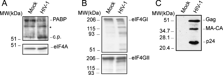
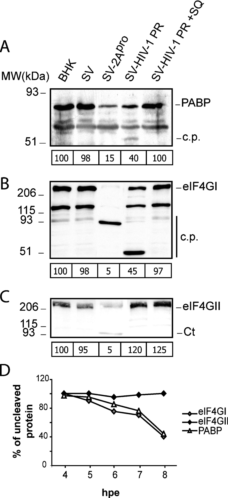
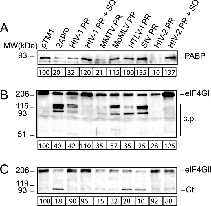
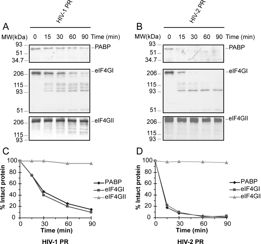
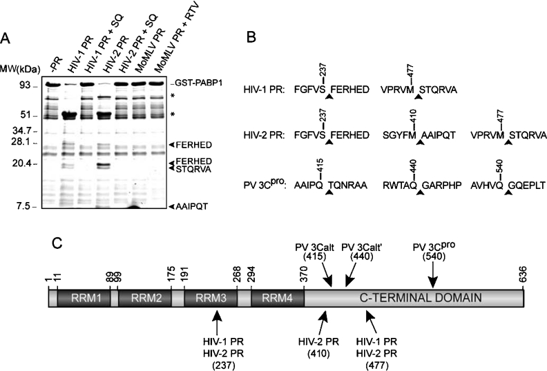
The PABP [poly(A)-binding protein] is able to interact with the 3' poly(A) tail of eukaryotic mRNA, promoting its translation. Cleavage of PABP by viral proteases encoded by several picornaviruses and caliciviruses plays a role in the abrogation of cellular protein synthesis. We report that infection of MT-2 cells with HIV-1 leads to efficient proteolysis of PABP. Analysis of PABP integrity was carried out in BHK-21 (baby-hamster kidney) and COS-7 cells upon individual expression of the protease from several members of the Retroviridae family, e.g. MoMLV (Moloney murine leukaemia virus), MMTV (mouse mammary tumour virus), HTLV-I (human T-cell leukaemia virus type I), SIV (simian immunodeficiency virus), HIV-1 and HIV-2. Moreover, protease activity against PABP was tested in a HeLa-cell-free system. Only MMTV, HIV-1 and HIV-2 proteases were able to cleave PABP in the absence of other viral proteins. Purified HIV-1 and HIV-2 proteases cleave PABP1 directly at positions 237 and 477, separating the two first RNA-recognition motifs from the C-terminal domain of PABP. An additional cleavage site located at position 410 was detected for HIV-2 protease. These findings indicate that some retroviruses may share with picornaviruses and caliciviruses the capacity to proteolyse PABP.


Figures

Figure 1
Figure 1. PABP cleavage in HIV-1-infected cells
(A) MT-2 cells were infected with HIV-1 (at an MOI of 1). At 3 dpi, the integrity of PABP was determined by Western blotting (upper panel; 10% gel). The corresponding Western blot against eIF4A is shown in the lower panel (10% gel). Mock, mock-infected cells; HIV-1, cells infected with HIV-1; c.p., cleavage product derived from PABP in infected cells; MW, molecular mass (sizes given in kDa). An unknown anti-PABP-reactive band is also denoted by an asterisk. (B) Western blots against eIF4GI (upper panel) and eIF4GII (lower panel) at 3 dpi (7.5% gel). (C) Western blot against the HIV-1 CA, p24 at 3 dpi (15% gel). The position of the intact initiation factors is also indicated in the respective panel.
Figure 2
Figure 2. Cleavage of PABP in cells electroporated with recombinant SVs that express heterologous proteases
(A) Cells were electroporated with transcription buffer (BHK), or wild-type SV (SV), SV-PR or SV-2A RNA and were incubated in the presence or absence of 12 μM SQ. At 8 hpe, cell extracts were analysed by Western blotting using specific antibodies against human PABP1 (10% gel). c.p., cleavage product derived from PABP in SV-PR-infected cells; MW, molecular mass (sizes given in kDa). The relative amount of intact PABP for each transfection experiment is indicated below each corresponding lane. (B) Detection of eIF4GI cleavage products by Western blotting using a mixture of antisera against its N- and C-terminal regions (7.5% gel). c.p., cleavage fragments. The amount of intact eIF4GI for each transfection experiment is indicated below each corresponding lane. (C) Detection of eIF4GII cleavage products by Western blotting using an antiserum against the C-terminal region (7.5% gel). Ct, C-terminal fragments of eIF4GII. The amount of intact eIF4GII for each transfection experiment is indicated below each lane. (D) Cleavage kinetics of PABP, eIF4GI and eIF4GII by HIV-1 protease. BHK-21 cells were electroporated as described in the Experimental section, and cell lysates were obtained at the indicated times. The values were obtained by densitometric scanning of the corresponding intact protein band at each time indicated.
Figure 3
Figure 3. Cleavage of PABP, eIF4GI and eIF4GII in transfected cells
(A) COS-7 cells were transfected with the empty pTM1 vector or with pTM1 carrying inserts containing the protease gene of several retroviruses or poliovirus 2Apro. At 16 hpt, equal amounts of protein extract were loaded in a polyacrylamide gel and analysed by Western blotting using a monoclonal antibody against PABP (10% gel). Data are referred to control experiments carried out with the empty pTM1 vector, and the values were obtained by densitometric scanning of the protein band of approx. 70 kDa corresponding to PABP. (B) Western blot against eIF4GI (7.5% gel). (C) Western blot against eIF4GII (7.5% gel). The estimated amount of intact protein in cell extracts obtained from transfections with different constructs is indicated below each panel. MW, molecular mass (sizes are given in kDa); c.p., cleavage fragments; Ct, C-terminal fragments of eIF4GII.
Figure 4
Figure 4. Cleavage of PABP in vitro by purified recombinant HIV-1, HIV-2 and MoMLV proteases
Crude HeLa S10 extracts (50 μg) were incubated with 3 ng/μl of each recombinant protease in a total volume of 20 μl for 90 min and subjected to Western blotting analysis by using: (A) a monoclonal antibody against PABP (10% gel); (B) a polyclonal antiserum against the 100 kDa subunit of eIF3 (top panel; 10% gel), an antibody against eIF4A (middle panel; 15% gel) or a monoclonal antibody raised against eIF4E (bottom panel; 15% gel). MW, molecular mass (sizes given in kDa).
Figure 5
Figure 5. Cleavage kinetics of PABP, eIF4GI and eIF4GII in vitro by purified recombinant HIV-1 and HIV-2 proteases
Crude HeLa S10 extracts (50 μg) were incubated with 3 ng/μl HIV-1 protease (A) or HIV-2 protease (B) in a total volume of 20 μl during the indicated times and subjected to Western blot analysis by using a monoclonal antibody against PABP (top panel; 10% gel), a mixture of antisera against N-terminal and C-terminal regions of eIF4GI (middle panel; 7.5% gel) or an antiserum against the C-terminal region of eIF4GII (bottom panel; 7.5% gel). Degradation kinetics of PABP, eIF4GI and eIF4GII in HeLa cell extracts incubated with HIV-1 protease (C) and HIV-2 protease (D). Determinations were obtained by densitometric scanning of the corresponding intact protein band at the indicated times. MW, molecular mass (sizes given in kDa).
Figure 6
Figure 6. Identification of HIV-1 protease and HIV-2 protease cleavage sites on PABP1
(A) Recombinant GST–PABP1 protein (5 μg) was incubated with 100 ng of recombinant HIV-1 protease, HIV-2 protease or MoMLV protease in a total volume of 20 μl as described in the Experimental section. Cleavage products were separated by SDS/17% PAGE, and the hydrolysis fragments were stained with Coomassie Brilliant Blue. The sequenced polypeptides are indicated with an arrowhead. Bands corresponding to GST N-terminal polypeptides are denoted by asterisks. MW, molecular mass (sizes given in kDa). (B) Amino acid sequence of the PABP1 cleavage sites identified using HIV-1 and HIV-2 proteases. (C) Diagram showing the functional domains found in PABP1 based on the available data, including the position of mapped cleavage sites for HIV-1 and HIV-2 proteases. The RRMs and the regions involved in the binding to other translation factors are shown. Sequence numbering refers to the human PABP1 isoform. RTV, ritonavir.

Comment in

References

    1. Hentze M. W. eIF4G: a multipurpose ribosome adapter? Science. 1997;275:500–501. - PubMed
    1. Jacobson A., Favreau M. Possible involvement of poly(A) in protein synthesis. Nucleic Acids Res. 1983;11:6353–6368. - PMC - PubMed
    1. Jacobson A. Poly(A) metabolism and translation: the closed loop model. In: Hershey J. W. B., Mathews M. B., Sonenberg N., editors. Translational Control. Cold Spring Harbor: Cold Spring Harbor Laboratory Press; 1996. pp. 451–480.
    1. Borman A. M., Michel Y. M., Kean K. M. Biochemical characterisation of cap-poly(A) synergy in rabbit reticulocyte lysates: the eIF4G–PABP interaction increases the functional affinity of eIF4E for the capped mRNA 5′-end. Nucleic Acids Res. 2000;28:4068–4075. - PMC - PubMed
    1. Tarun S. Z., Jr, Wells S. E., Deardorff J. A., Sachs A. B. Translation initiation factor eIF4G mediates in vitro poly(A) tail-dependent translation. Proc. Natl. Acad. Sci. U.S.A. 1997;94:9046–9051. - PMC - PubMed

Publication types

MeSH terms