

Mitochondrial aconitase and citrate metabolism in malignant and nonmalignant human prostate tissues
- PMID: 16595004
- PMCID: PMC1484490
- DOI: 10.1186/1476-4598-5-14
Mitochondrial aconitase and citrate metabolism in malignant and nonmalignant human prostate tissues
Abstract
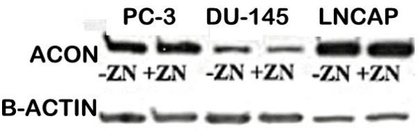
Background: In prostate cancer, normal citrate-producing glandular secretory epithelial cells undergo a metabolic transformation to malignant citrate-oxidizing cells. m-Aconitase is the critical step involved in this altered citrate metabolism that is essential to prostate malignancy. The limiting m-aconitase activity in prostate epithelial cells could be the result of a decreased level of m-aconitase enzyme and/or the inhibition of existing m-aconitase. Earlier studies identified zinc as an inhibitor of m-aconitase activity in prostate cells; and that the depletion of zinc in malignant cells is an important factor in this metabolic transformation. However, a possibility remains that an altered expression and level of m-aconitase enzyme might also be involved in this metabolic transformation. To address this issue, the in situ level of m-aconitase enzyme was determined by immunohistochemical analysis of prostate cancer tissue sections and malignant prostate cell lines.
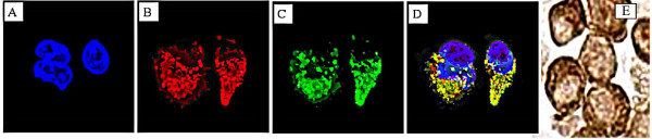
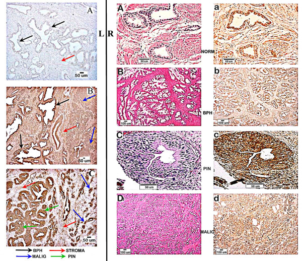
Results: The immunocytochemical procedure successfully identified the presence of m-aconitase localized in the mitochondrial compartment in PC-3, LNCaP, and DU-145 malignant prostate cell lines. The examination of prostate tissue sections from prostate cancer subjects demonstrated that m-aconitase enzyme is present in the glandular epithelium of normal glands, hyperplastic glands, adenocrcinomatous glands, and prostatic intraepithelial neoplastic foci. Quantitative analysis of the relative level of m-aconitase in the glandular epithelium of citrate-producing adenomatous glands versus the citrate-oxidizing adenocarcinomatous glands revealed no significant difference in m-aconitase enzyme levels. This is in contrast to the down-regulation of ZIP1 zinc transporter in the malignant glands versus hyperplastic glands that exists in the same tissue samples.
Conclusion: The results demonstrate the existence of m-aconitase enzyme in the citrate-producing glandular epithelial cells; so that deficient m-aconitase enzyme is not associated with the limiting m-aconitase activity that prevents citrate oxidation in these cells. The level of m-aconitase is maintained in the malignant cells; so that an altered enzyme level is not associated with the increased m-aconitase activity. Consequently, the elevated zinc level that inhibits m-aconitase enzyme is responsible for the impaired citrate oxidation in normal and hyperplastic prostate glandular epithelial cells. Moreover, the down-regulation of ZIP1 zinc transporter and corresponding depletion of zinc results in the increase in the activity of the existing m-aconitase activity in the malignant prostate cells. The studies now define the mechanism for the metabolic transformation that characterizes the essential transition of normal citrate-producing epithelial cells to malignant citrate-oxidizing cells.
Figures
References
-
- LC C, RB F. The metabolism of prostate malignancy: Insights into the pathogenesis of prostate cancer and new approaches for its diagnosis and treatment. Oncology Spectrums. 2001;2:452–457.
-
- Costello L, Franklin RB, Kurhanewicz J. The metabolic diagnosis of prostate by magnetic resonance spectroscopy. 2 nd. Vol. 3. 2002. pp. 167–177.
-
- Costello LC, Franklin RB. - Concepts of citrate production and secretion by prostate. 1. Metabolic relationships - PubMed
Publication types
MeSH terms
Substances
Grants and funding
LinkOut - more resources
Full Text Sources
Other Literature Sources
Medical
