. 2006 Mar-Apr;41(2):77-102.
doi: 10.1080/10409230600602634.

Ribonuclease P: the evolution of an ancient RNA enzyme

Affiliations

Ribonuclease P: the evolution of an ancient RNA enzyme

Scott C Walker et al. Crit Rev Biochem Mol Biol. 2006 Mar-Apr.

Abstract

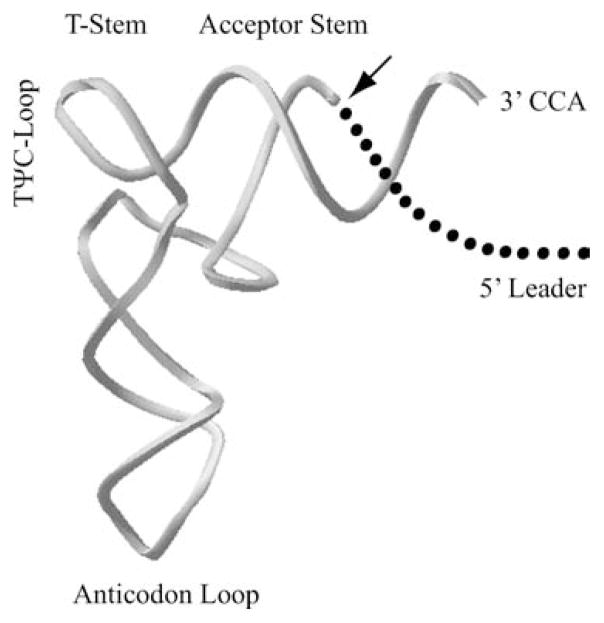
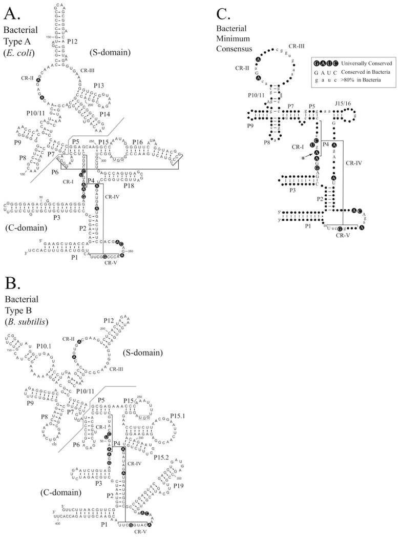
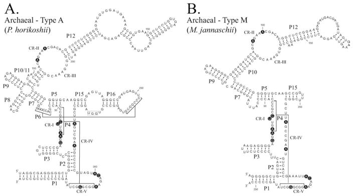
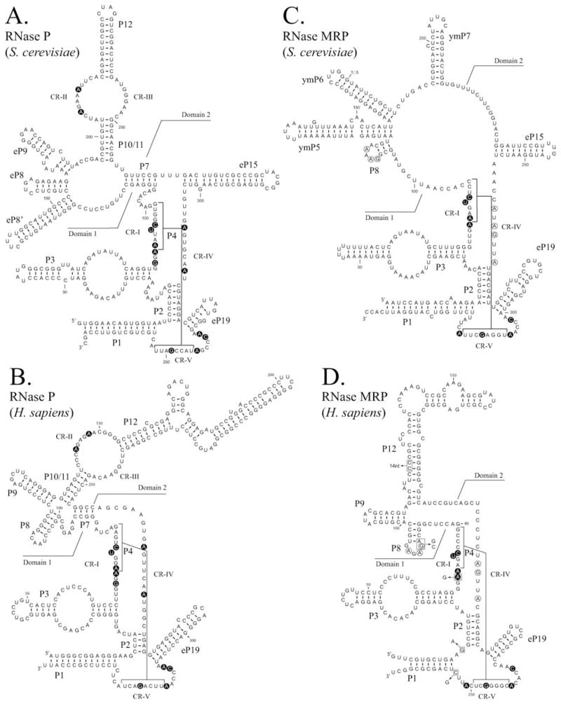
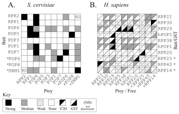
Ribonuclease P (RNase P) is an ancient and essential endonuclease that catalyses the cleavage of the 5' leader sequence from precursor tRNAs (pre-tRNAs). The enzyme is one of only two ribozymes which can be found in all kingdoms of life (Bacteria, Archaea, and Eukarya). Most forms of RNase P are ribonucleoproteins; the bacterial enzyme possesses a single catalytic RNA and one small protein. However, in archaea and eukarya the enzyme has evolved an increasingly more complex protein composition, whilst retaining a structurally related RNA subunit. The reasons for this additional complexity are not currently understood. Furthermore, the eukaryotic RNase P has evolved into several different enzymes including a nuclear activity, organellar activities, and the evolution of a distinct but closely related enzyme, RNase MRP, which has different substrate specificities, primarily involved in ribosomal RNA biogenesis. Here we examine the relationship between the bacterial and archaeal RNase P with the eukaryotic enzyme, and summarize recent progress in characterizing the archaeal enzyme. We review current information regarding the nuclear RNase P and RNase MRP enzymes in the eukaryotes, focusing on the relationship between these enzymes by examining their composition, structure and functions.


Figures

FIGURE 1
FIGURE 1
RNase P cleaves the 5′ leader from precursor tRNAs (pre-tRNAs). The site of cleavage by RNase P is represented with an arrow. Structures required for substrate recognition and cleavage by RNase P, the acceptor stem and T-stem-loop, are indicated.
FIGURE 2
FIGURE 2
Comparison of Type A and Type B bacterial RNase P RNAs. The secondary structures of RNase P RNAs derived by phylogenetic comparative sequence analysis are shown (Haas and Brown, 1998). (A). The secondary structure of the E. coli RNase P RNA (Type A). (B). The secondary structure of the B. subtilis RNase P RNA (Type B). Helices are numbered as paired regions (e.g. P1), other helices are indicated by lines (e.g. P4 and P6). Regions of the RNA implicated in substrate binding the “specificity domain” (S-domain) and catalytic activity the “catalytic domain” (C-domain) are separated by a line. A boxed region within the loop of P15 indicates a binding site for the 3′CCA of the pre-tRNA substrate, which is present in many bacterial RNase P RNAs. Nucleotides conserved in all known RNase P RNAs are shown in bold background. (C). Consensus structure showing elements common to all known bacterial RNase P RNAs (figure adapted from Marquez et al., 2005). The highlighted conserved adenine has been shown to be extrahelical in the structure of the Bacillus stearothermophilus RNase P RNA (Kazantsev et al., 2005). In this structure, the adjacent conserved adenosine forms the corresponding base pair within the P4 helix.
FIGURE 3
FIGURE 3
Comparision of Type A and Type M archaeal RNase P RNAs. The secondary structures of RNase P RNAs derived by phylogenetic comparative sequence analysis are shown (Haas et al., 1996). (A). The secondary structure of the Pyrococcus horikoshii OT3 RNase P RNA (Type A). (B). The secondary structure of the Methanococcus jannaschii RNase P RNA (Type M). Structures are represented in a similar manner to Figure 2. Nucleotides conserved in all known RNase P RNAs are shown in bold background.
FIGURE 4
FIGURE 4
Summary of protein-protein interactions detected in archaeal systems. Data from previously published yeast two-hybrid studies are summarized. Tables are drawn such that homologous proteins are represented in similar positions in both tables, starting from the top left corner. The interactions are indicated as described in the original text. (A). Interactions demonstrated in protein components of the Pyrococcus horikoshii OT3 RNase P (Kifusa et al., 2005). (B). Interactions demonstrated in protein components of the Methanothermobacter thermoautotrophicus RNase P (Hall and Brown, 2004).
FIGURE 5
FIGURE 5
Comparison of eukaryotic nuclear RNase P RNAs and RNase MRP RNAs. Structures are represented in a similar manner to Figure 2. RNase P RNAs (A) and (B) and RNase MRP RNAs (C) and (D) are shown for Saccharomyces cerevisiae and Homo sapiens respectively. RNase MRP structures are based on previous structural data and are updated according to (Piccinelli et al., 2005). Nucleotides conserved in all known RNase P RNAs or RNase MRP RNAs are shown in bold background. Line boundaries do not precisely define domains, but draw attention to regions of similarity. In both RNase P and RNase MRP RNAs domain 1 shows similarity to the bacterial “catalytic domain” (C-domain). In the MRP RNAs the conserved region (CR-IV) has the general consensus (5′-ANAGNNA-3′) and the P8 loop has the general consensus (5′-GARAR-3′) [R = purine], these highly conserved nucleotides are circled. Mutations and insertions within the mature RNase MRP RNA that are known to be involved in the human diseases cartilage-hair hypoplasia (CHH) and anauxetic dysplasia (Ridanpaa et al., 2002; Thiel et al., 2005), are indicated with boxes in (D).
FIGURE 6
FIGURE 6
Summary of protein-protein interactions detected in eukaryotic systems. Data from previously published yeast two-hybrid (Y2H) and glutathione-S-transferase (GST) pull down studies are summarized. Tables are drawn such that homologous proteins are represented in similar positions in both tables, starting from the top left corner, and are arranged to allow a direct comparison with Figure 4. Proteins that have no identified homologue between yeast and humans are indicated with an asterisk. (A). Interactions demonstrated in the yeast system (Saccharomyces cerevisiae). Y2H interactions are shown as described in the original text (Houser-Scott et al., 2002). (B). Interactions demonstrated in the vertebrate system (Homo sapiens) interactions are shown as described in the original text (Jiang and Altman, 2001; Welting et al., 2004).

Similar articles

Cited by

References

    1. Alifano P, Rivellini F, Piscitelli C, Arraiano CM, Bruni CB, Carlomagno MS. Ribonuclease E provides substrates for ribonuclease P-dependent processing of a polycistronic mRNA. Genes Dev. 1994;8(24):3021. - PubMed
    1. Altman S, Wesolowski D, Guerrier-Takada C, Li Y. RNase P cleaves transient structures in some riboswitches. Proc Natl Acad Sci USA. 2005;102(32):11284. - PMC - PubMed
    1. Altman S, Wesolowski D, Puranam RS. Nucleotide sequences of the RNA subunit of RNase P from several mammals. Genomics. 1993;18(2):418. - PubMed
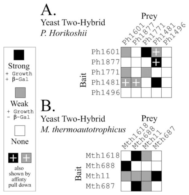
    1. Andrews AJ, Hall TA, Brown JW. Characterization of RNase P holoenzymes from Methanococcus jannaschii and Methanothermobacter thermoautotrophicus. Biol Chem. 2001;382(8):1171. - PubMed
    1. Aravind L, Iyer LM, Anantharaman V. The two faces of Alba: the evolutionary connection between proteins participating in chromatin structure and RNA metabolism. Genome Biol. 2003;4(10):R64. - PMC - PubMed

Publication types

LinkOut - more resources