. 2006 Apr 5;26(14):3685-96.
doi: 10.1523/JNEUROSCI.0414-06.2006.

Inclusion body formation and neurodegeneration are parkin independent in a mouse model of alpha-synucleinopathy

Affiliations

Inclusion body formation and neurodegeneration are parkin independent in a mouse model of alpha-synucleinopathy

Rainer von Coelln et al. J Neurosci. .

Abstract

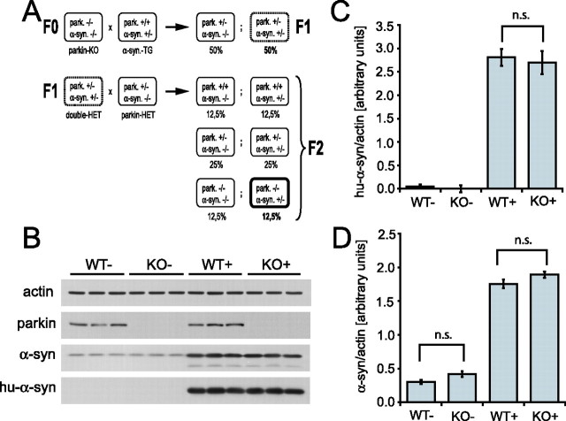
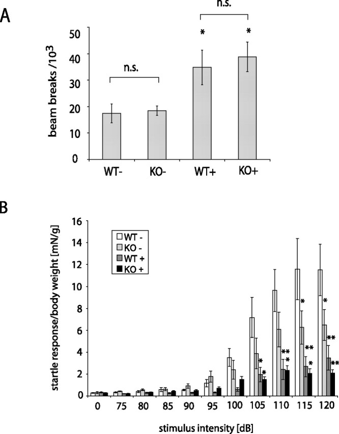
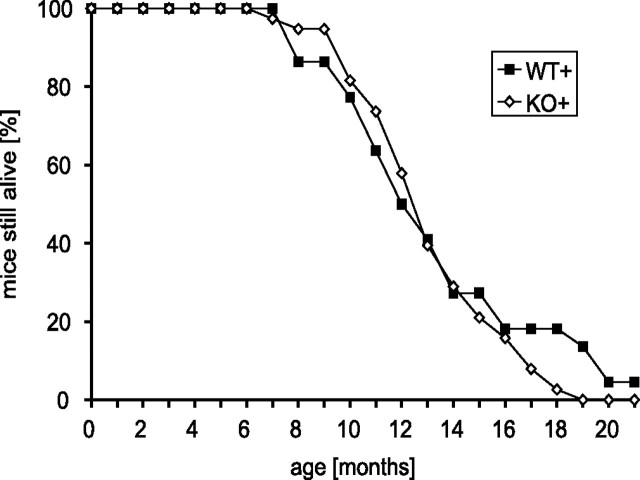
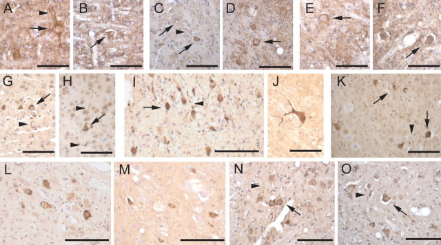
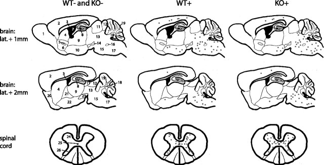
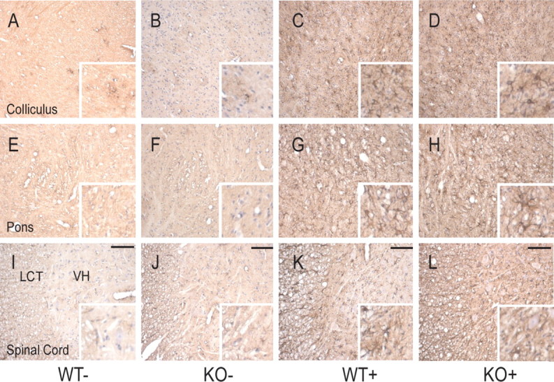
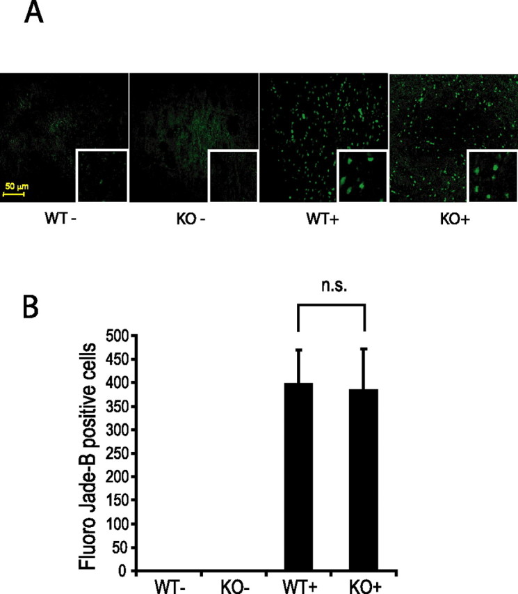
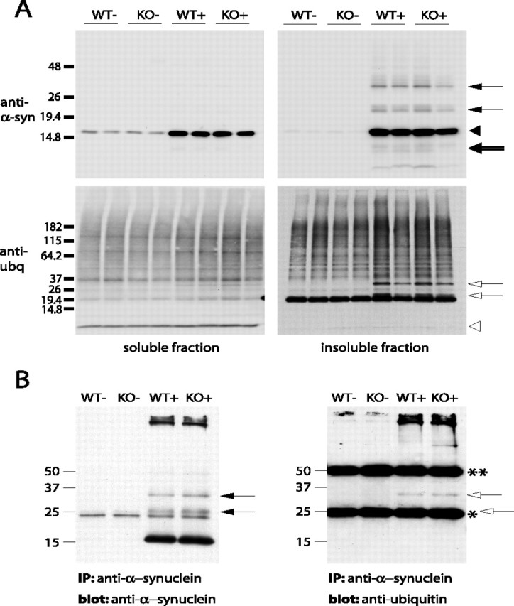
Mutations in the genes coding for alpha-synuclein and parkin cause autosomal-dominant and autosomal-recessive forms of Parkinson's disease (PD), respectively. Alpha-synuclein is a major component of Lewy bodies, the proteinaceous cytoplasmic inclusions that are the pathological hallmark of idiopathic PD. Lewy bodies appear to be absent in cases of familial PD associated with mutated forms of parkin. Parkin is an ubiquitin E3 ligase, and it may be involved in the processing and/or degradation of alpha-synuclein, as well as in the formation of Lewy bodies. Here we report the behavioral, biochemical, and histochemical characterization of double-mutant mice overexpressing mutant human A53T alpha-synuclein on a parkin null background. We find that the absence of parkin does not have an impact on the onset and progression of the lethal phenotype induced by overexpression of human A53T alpha-synuclein. Furthermore, all major behavioral, biochemical, and morphological characteristics of A53T alpha-synuclein-overexpressing mice are not altered in parkin null alpha-synuclein-overexpressing double-mutant mice. Our results demonstrate that mutant alpha-synuclein induces neurodegeneration independent of parkin-mediated ubiquitin E3 ligase activity in nondopaminergic systems and suggest that PD caused by alpha-synuclein and parkin mutations may occur via independent mechanisms.


Figures

Figure 1.
Figure 1.
Absence of parkin does not change steady-state levels of α-synuclein. A, Breeding strategy to generate parkin null α-synuclein-overexpressing double-mutant mice. F0, F1, and F2 represent the generations of crossbreeding. Percentages represent the expected frequency of each genotype in a sufficiently large number of litters. Het, Heterozygous; TG, transgenic, i.e., A53T α-synuclein-overexpressing mouse. B, Immunoblot analysis of NP 40-soluble lysates from cerebral cortex of 6-month-old mice with or without parkin and with or without overexpressed human A53T α-synuclein was performed using antibodies that recognize both mouse and human α-synuclein (α-syn) and antibodies that recognize only overexpressed human α-synuclein (hu-α-syn). An antibody to actin was used as a loading control, and an antibody to parkin was used to verify the absence of parkin. The expression level of α-synuclein (normalized to actin) was quantified using optical densitometry for overexpressed human A53T α-synuclein (C) and total (mouse and human) α-synuclein (D). Data are expressed as mean ± SEM (n = 3). Statistical analysis was performed by ANOVA, followed by the Student–Newman–Keuls test. n.s., Not statistically significant.
Figure 2.
Figure 2.
Behavioral deficits in mice overexpressing human A53T α-synuclein are not altered by the absence of parkin. A, The Open-field test (n = 6–8) shows hyperactivity of transgenic mice in the presence and absence of parkin. B, Acoustic startle analysis (n = 5–7) reveals reduced startle in parkin null mice and a reduction of the startle response in A53T α-synuclein-overexpressing mice, as well as in double-mutant mice. Data are expressed as mean ± SEM. Statistical analysis was performed by ANOVA, followed by the Student–Newman–Keuls test (*p < 0.05; **p < 0.01). n.s., Not statistically significant.
Figure 3.
Figure 3.
Progression of the lethal phenotype induced by overexpression of human A53T α-synuclein is independent of parkin. Survival curves of transgenic mice on a parkin WT and KO background, respectively, were generated by following cohorts of both genotypes (WT+, n = 22; KO+, n = 38) until mice had to be killed because of terminal disease.
Figure 4.
Figure 4.
Abnormal accumulation of human α-synuclein and ubiquitin in transgenic mice overexpressing human A53T α-synuclein is not affected by a lack of parkin expression. Paraffin-embedded brain sections from parkin WT/A53T α-synuclein transgenic mice (A, C, E, G, L, N) or parkin null A53T α-synuclein transgenic mice (B, D, F, H, I–K, M, O) were immunolabeled for human α-synuclein (A–F) or ubiquitin (G–O). Shown are representative images from pons (A, B, I, J), deep cerebellar nuclei (C, D, K), spinal cord (E, F, N, O) cortex (G, H), and red nucleus (L, M). Abnormal accumulation of human α-synuclein or ubiquitin in cell bodies (arrows) and processes (arrowheads) associated with the expression of mutant human A53T α-synuclein were not significantly affected by the lack of parkin expression. Scale bars: A–H, J, K, O, 50 μm; I, L–N, 100 μm.
Figure 5.
Figure 5.
Anatomical distribution of pathological protein inclusions in α-synuclein transgenic mice is unaltered by the absence of parkin. Sagittal brain sections and transversal spinal cord sections of mice killed at the terminal stage of α-synuclein-induced disease (or age-matched for WT− and KO−) were analyzed regarding the distribution of ubiquitin-immunoreactive protein inclusions. Each black dot represents one immunoreactive neuronal cell on a typical brain section of an affected animal. 1, Olfactory bulb; 2, cerebral cortex; 3, corpus callosum; 4, striatum; 5, anterior commissure; 6, nucleus accumbens; 7, third ventricle; 8, hippocampus; 9, thalamus; 10, hypothalamus; 11, superior colliculus; 12, inferior colliculus; 13, mesencephalon; 14, substantia nigra, pars compacta; 15, pons; 16, facial nerve; 17, medulla oblongata; 18, deep cerebellar nuclei; 19, cerebellar cortex; 20, olfactory tubercle; 21, globus pallidus; 22, amygdala; 23, substantia nigra, pars reticulata; 24, ventral horn; 25, central canal; 26, dorsal horn.
Figure 6.
Figure 6.
The pathology-associated astrocytic response in A53T α-synuclein transgenic mice is not affected by the lack of parkin expression in parkin KO/α-synuclein transgenic double-mutant mice. Paraffin-embedded brain sections were immunostained for GFAP. Representative sections from colliculus (A–D), pons (E, F), and cervical spinal cord (I–L) are shown in overview and detail (inset). Scale bars, 50 μm. A normal pattern of astrocytic staining (with sparse GFAP-positive cells) is observed in nontransgenic mice with or without parkin (A, B, E, F, I, J). In clinically affected A53T α-synuclein transgenic mice (C, G, K), GFAP staining reveals a significant astrocytic response. The pattern of GFAP staining is not obviously affected by the lack of parkin expression (D, H, L). Note that, in spinal cord, activation of astrocytes is seen in both the lateral column tracts (LCT) and in ventral horn (VH).
Figure 7.
Figure 7.
Neurodegeneration in A53T α-synuclein transgenic mice as visualized by Fluoro Jade B staining is unaltered by the absence of parkin. Sagittal brain sections at the level of brainstem of mice killed at the terminal stage of α-synuclein-induced disease (or age-matched for WT− and KO−) were analyzed regarding the quantity and distribution of Fluoro Jade B-positive cells. A, Fluoro Jade B-positive cells are abundant in α-synuclein transgenic mice regardless of the presence of parkin. B, Quantification of Fluoro Jade B-positive cells. Data are expressed as mean ± SEM (n = 4). Statistical analysis was performed by ANOVA, followed by the Student–Newman–Keuls test. Scale bar, 50 μm. n.s., Not statistically significant.
Figure 8.
Figure 8.
Accumulation of insoluble α-synuclein, proteolytic processing, and ubiquitination of α-synuclein are parkin independent. Brainstem lysates of terminally ill (or age-matched control) mice were separated into non-ionic detergent-soluble and -insoluble fractions. Immunoblotting was performed using antibodies against α-synuclein and ubiquitin of both soluble and insoluble fraction (A) and of immunoprecipitated (IP) α-synuclein (B). Filled arrows, Mono- and di-ubiquitinated α-synuclein species. Filled arrowhead, α-Synuclein monomer, unmodified. Double-lined arrow, Proteolytically processed α-synuclein fragments. Open arrows, Bands corresponding to the mono- and di-ubiquitinated α-synuclein species. Open arrowhead, Ubiquitin monomer. Dotted open arrow, Mono-ubiquitinated α-synuclein species covered by the IgG light chain after immunoprecipitation. *IgG light chain; **IgG heavy chain. Molecular weight markers are indicated.

References

    1. Baba M, Nakajo S, Tu PH, Tomita T, Nakaya K, Lee VM, Trojanowski JQ, Iwatsubo T (1998). Aggregation of alpha-synuclein in Lewy bodies of sporadic Parkinson's disease and dementia with Lewy bodies. Am J Pathol 152:879–884. - PMC - PubMed
    1. Bennett MC, Bishop JF, Leng Y, Chock PB, Chase TN, Mouradian MM (1999). Degradation of alpha-synuclein by proteasome. J Biol Chem 274:33855–33858. - PubMed
    1. Braak H, Del Tredici K, Rub U, de Vos RA, Jansen Steur EN, Braak E (2003). Staging of brain pathology related to sporadic Parkinson's disease. Neurobiol Aging 24:197–211. - PubMed
    1. Chung KK, Zhang Y, Lim KL, Tanaka Y, Huang H, Gao J, Ross CA, Dawson VL, Dawson TM (2001). Parkin ubiquitinates the alpha-synuclein-interacting protein, synphilin-1: implications for Lewy-body formation in Parkinson disease. Nat Med 7:1144–1150. - PubMed
    1. Chung KK, Thomas B, Li X, Pletnikova O, Troncoso JC, Marsh L, Dawson VL, Dawson TM (2004). S-nitrosylation of parkin regulates ubiquitination and compromises parkin's protective function. Science 304:1328–1331. - PubMed

Publication types

MeSH terms