. 2006 Apr 5;26(14):3798-804.
doi: 10.1523/JNEUROSCI.5338-05.2006.

Inhibition of apoptosis by P2Y2 receptor activation: novel pathways for neuronal survival

Affiliations

Inhibition of apoptosis by P2Y2 receptor activation: novel pathways for neuronal survival

David B Arthur et al. J Neurosci. .

Abstract

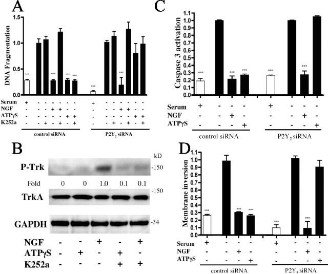
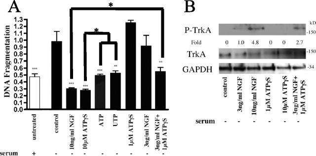
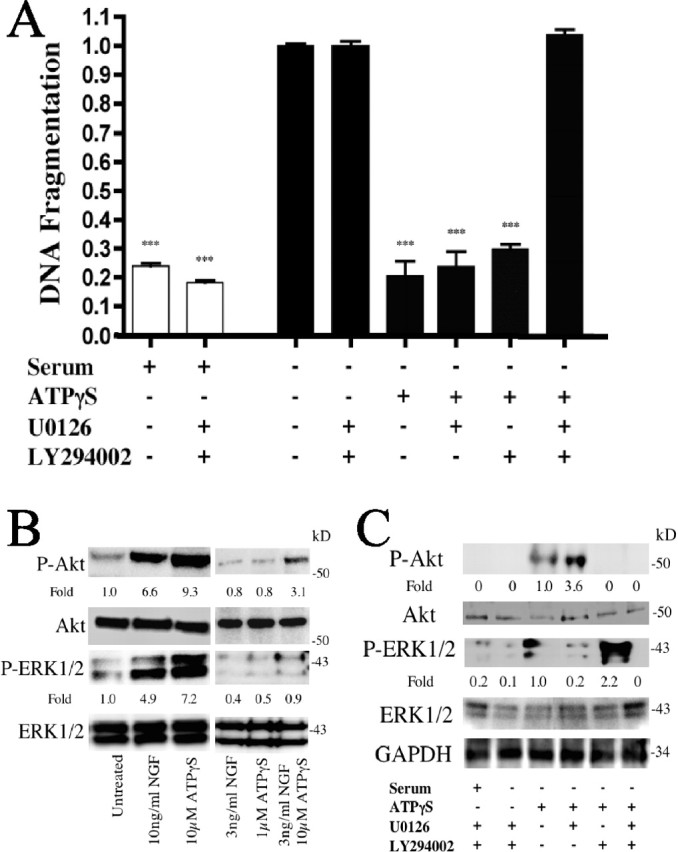
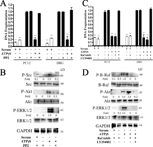
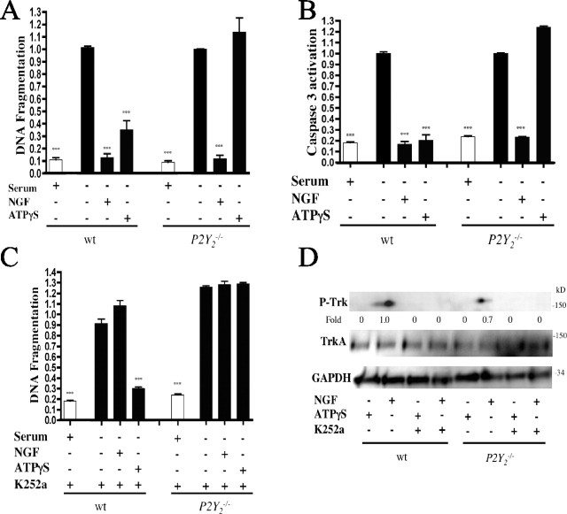
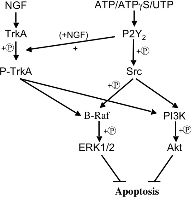
Cell survival is an essential function in the development and maintenance of the nervous system. We demonstrate here a previously unappreciated role for extracellular nucleotide signaling through the P2Y2 receptor in the survival of neurons: PC12 (pheochromocytoma 12) cells and dorsal root ganglion neurons are protected from serum starvation-induced apoptosis by ATP, UTP, and ATPgammaS, an effect mediated via P2Y2 receptors, as demonstrated by small interfering RNA and genetic knock-out models. This protection occurs independently of neurophin signaling but requires Src activation of ERK (extracellular signal-regulated kinase) and Akt. Moreover, ATPgammaS and NGF act synergistically to enhance neuronal survival through enhanced TrkA signaling. The results, which define a novel mechanism for inhibition of apoptosis, implicate parallel, interacting systems--extracellular nucleotides/P2Y2 receptors and neurotrophin/TrkA--to sustain neuronal survival.


Figures

Figure 1.
Figure 1.
ATPγS inhibits serum starvation-induced PC12 apoptosis via P2Y2 receptors independent of NGF/TrkA signaling. PC12 cells were transfected with P2Y2 siRNA and then grown for 12 h in the presence or absence of serum, ATPγS (10 μm), NGF (10 ng/ml), and/or K252a (10 nm) and analyzed for apoptosis by quantitation of DNA fragmentation (A). P2Y2 receptor expression in cells transfected with P2Y2 siRNA decreased by >70% versus cells transfected with a scrambled siRNA sequence. Treatment with the P2Y2-targeted siRNAs failed to alter serum starvation-induced apoptosis. An immunoblot of PC12 cells treated as in A and probed for TrkA activation is shown (B). Densitometry was measured as P-TrkA/TrkA and normalized to NGF treatment alone. PC12 cells transfected with P2Y2 siRNA and serum-starved for 12 h with the indicated treatments were analyzed for apoptosis by caspase 3 activation/expression (C) and an assay for plasma membrane inversion (D). ***p < 0.001 versus serum starvation alone, with values normalized to serum starvation alone. Error bars indicate SE.
Figure 2.
Figure 2.
ATPγS/NGF interact to enhance inhibition of apoptosis. Serum-starved PC12 cells were treated with ATP (100 μm), UTP (100 μm), NGF, and/or ATPγS at the indicated concentrations. Apoptosis was quantitated using DNA fragmentation (A). An immunoblot analysis of cells treated with NGF (10 or 3 ng/ml), ATPγS (10 or 1 μm), or 3 ng/ml NGF together with 1 μm ATPγS and probed for TrkA activation is shown (B). Densitometry was measured as P-TrkA/TrkA and normalized to 3 ng/ml NGF treatment alone. *p < 0.05, **p < 0.01, ***p < 0.001 versus serum starvation alone, with values normalized to serum starvation alone. Error bars indicate SE.
Figure 3.
Figure 3.
ATPγS inhibits apoptosis via activation of ERK1/2 and Akt. Serum-starved PC12 cells were treated with U0126 (10 μm) or LY294002 (10 μm), and the indicated treatments were analyzed for apoptosis by quantitation of DNA fragmentation (A). An immunoblot of serum-starved PC12 cells left untreated, treated with 10 ng/ml NGF, 10 μm ATPγS, or as indicated and probed for activated Akt and ERK1/2 is shown (B). Densitometry was measured as P-Akt/Akt and P-ERK1/2/ERK1/2 and normalized to untreated. Immunoblot of serum-starved PC12 cells treated as in A and probed for Akt and ERK1/2 activation is shown (C). Densitometry was measured as P-Akt/Akt and P-ERK1/2/ERK1/2 and normalized to ATPγS-treated alone. ***p < 0.001 versus serum starvation alone, with values normalized to serum starvation alone. Error bars indicate SE.
Figure 4.
Figure 4.
ATPγS inhibits apoptosis via Src activation of Akt and B-Raf-mediated ERK1/2 activation. Serum-starved PC12 cells and DRG neurons were treated with 10 μm PP2 and 10 μm ATPγS. Cells were analyzed for apoptosis by quantitation of DNA fragmentation (A). An immunoblot of PC12 cells treated as in A and probed for Src, Akt, and ERK activation is shown (B). Densitometry was measured as P-Src/Src, P-Akt/Akt, and P-ERK1/2/ERK1/2 and normalized to ATPγS-treated alone. Serum-starved PC12 cells and DRG neurons were treated with a Raf kinase inhibitor (50 nm) and/or an Akt inhibitor (LY294002; 10 μm). Cells were analyzed for apoptosis by quantitation of DNA fragmentation (C). An immunoblot of PC12 cells treated as in C and probed for B-Raf, Akt, and ERK activation is shown (D). Densitometry was measured as P-B-Raf/Raf, P-Akt/Akt, and P-ERK1/2/ERK1/2 and normalized to ATPγS-treated alone. ***p < 0.001 versus serum starvation alone, with values normalized to serum starvation alone. Error bars indicate SE.
Figure 5.
Figure 5.
ATPγS inhibits serum starvation-induced DRG apoptosis via P2Y2 independent of NGF/TrkA signaling. DRG neurons from wt and P2Y2−/− mice were serum-deprived for 12 h alone, with 10 ng/ml NGF, or with 10 μm ATPγS. Apoptosis was quantitated by DNA fragmentation (A) or caspase 3 expression (B). DRG neurons from wt and P2Y2−/− mice were serum-starved for 12 h in the presence of the TrkA inhibitor K252a (10 nm) and the indicated treatments (compare with A). Cells were lysed and analyzed for apoptosis by DNA fragmentation (C). An immunoblot analysis of DRG neurons treated as in C and probed for TrkA activation is shown (D). Densitometry was measured as P-TrkA/TrkA and normalized to NGF treatment alone. ***p < 0.001 versus serum starvation alone, values normalized to serum starvation alone. Error bars indicate SE.
Figure 6.
Figure 6.
Model of P2Y2-mediated inhibition of neuronal apoptosis. Agonists (e.g., ATP, ATPγS, or UTP) activate P2Y2 receptors leading to Src activation/phosphorylation (+P). Src activates/phosphorylates B-Raf and PI3K (phosphatidylinositol 3-kinase) leading to ERK1/2 and Akt activation/phosphorylation, respectively. Activation of ERK1/2 and Akt inhibits apoptosis. Activation of P2Y2 receptors in the presence of NGF increases TrkA activation/phosphorylation, thereby increasing the activation of ERK1/2 and Akt, resulting in inhibition of apoptosis via a NGF-dependent pathway.

References

    1. Akassoglou K (2005). Nerve growth factor-independent neuronal survival: a role for NO donors. Mol Pharmacol 68:952–955. - PubMed
    1. Arthur DB, Akassoglou K, Insel PA (2005). P2Y2 receptor activates nerve growth factor/TrkA receptor signaling to enhance neuronal differentiation. Proc Natl Acad Sci USA 102:19138–19143. - PMC - PubMed
    1. Bacon KB, Camp RD (1990). Interleukin (IL)-8-induced in vitro human lymphocyte migration is inhibited by cholera and pertussis toxins and inhibitors of protein kinase C. Biochem Biophys Res Commun 169:1099–1104. - PubMed
    1. Batistatou A, Greene LA (1993). Internucleosomal DNA cleavage and neuronal cell survival/death. J Cell Biol 122:523–532. - PMC - PubMed
    1. Berra E, Diaz-Meco MT, Moscat J (1998). The activation of p38 and apoptosis by the inhibition of Erk is antagonized by the phosphoinositide 3-kinase/Akt pathway. J Biol Chem 273:10792–10797. - PubMed

Publication types

LinkOut - more resources