. 2006 May;15(5):1142-52.
doi: 10.1110/ps.052045306. Epub 2006 Apr 5.

Structure and dynamics of the epidermal growth factor receptor C-terminal phosphorylation domain

Affiliations

Structure and dynamics of the epidermal growth factor receptor C-terminal phosphorylation domain

Nam Y Lee et al. Protein Sci. 2006 May.

Abstract

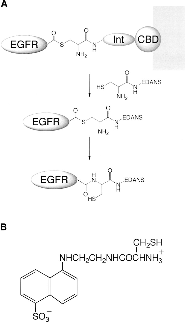
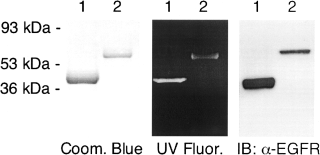
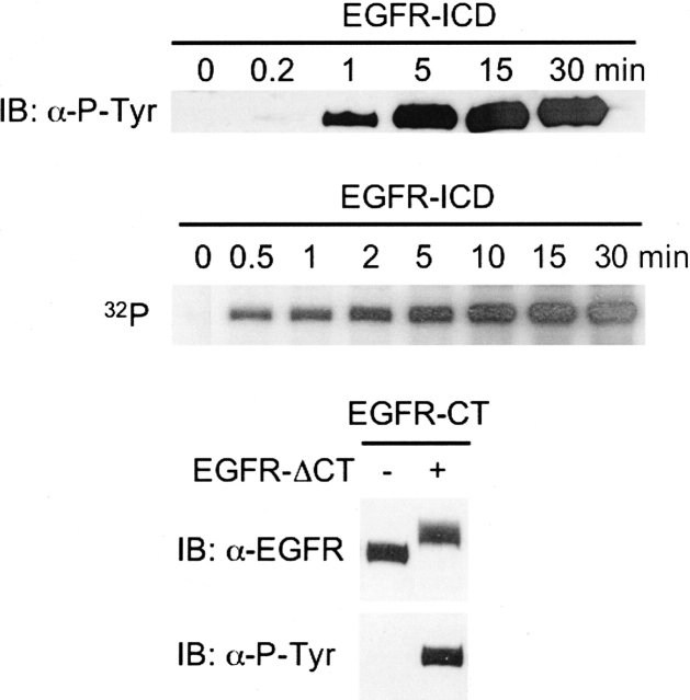
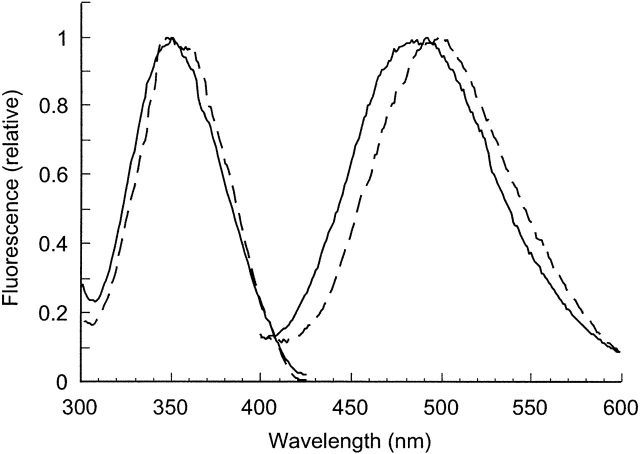
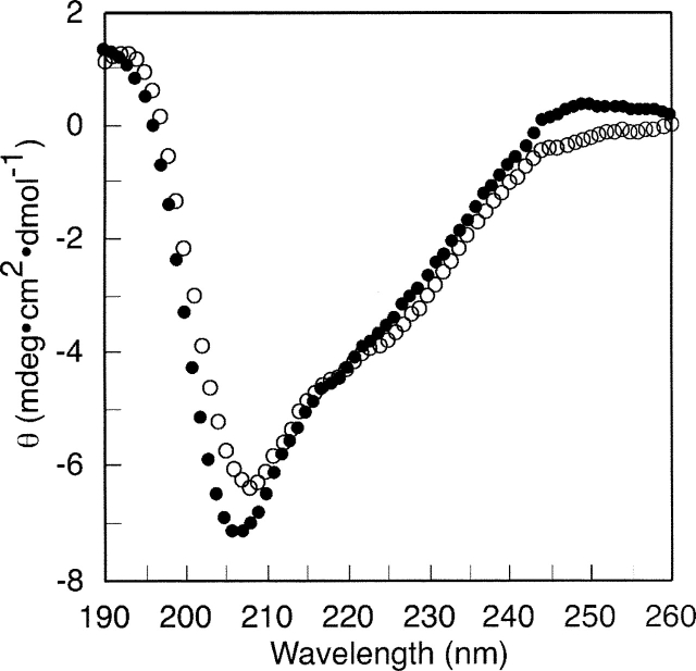
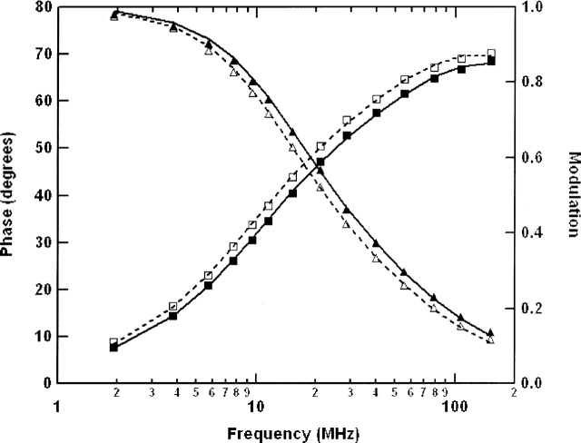
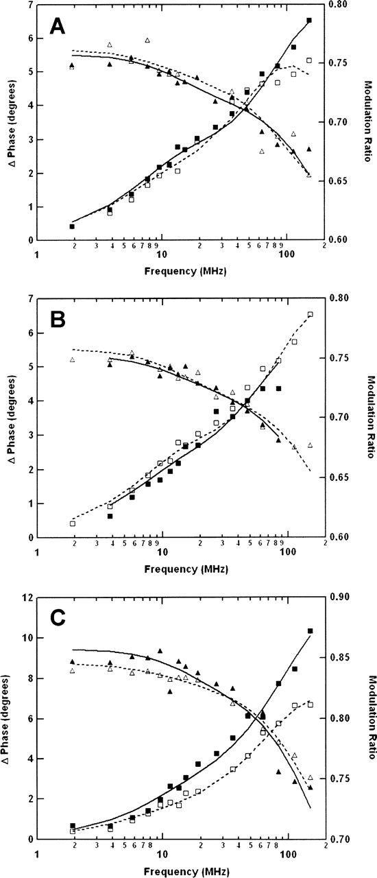
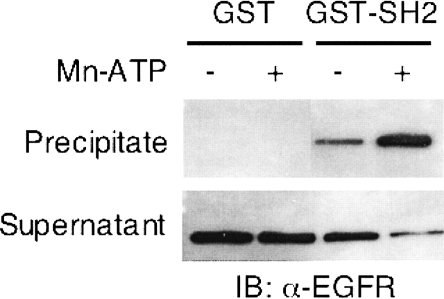
The C-terminal phosphorylation domain of the epidermal growth factor receptor is believed to regulate protein kinase activity as well as mediate the assembly of signal transduction complexes. The structure and dynamics of this proposed autoregulatory domain were examined by labeling the extreme C terminus of the EGFR intracellular domain (ICD) with an extrinsic fluorophore. Fluorescence anisotropy decay analysis of the nonphosphorylated EGFR-ICD yielded two rotational correlation times: a longer time, consistent with the global rotational motion of a 60- to 70-kDa protein with an elongated globular conformation, and a shorter time, presumably contributed by segmental motion near the fluorophore. A C-terminally truncated form of EGFR-ICD yielded a slow component consistent with the rotational motion of the 38-kDa kinase core. These findings suggested a structural arrangement of the EGFR-ICD in which the C-terminal phosphorylation domain interacts with the kinase core to move as an extended structure. A marked reduction in the larger correlation time of EGFR-ICD was observed upon its autophosphorylation. This dynamic component was faster than predicted for the globular motion of the 62-kDa EGFR-ICD, suggesting an increase in the mobility of the C-terminal domain and a likely displacement of this domain from the kinase core. The interaction between the SH2 domain of c-Src and the phosphorylated EGFR C-terminal domain was shown to impede its mobility. Circular dichroism spectroscopy indicated that the EGFR C-terminal domain possessed a significant level of secondary structure in the form of alpha-helices and beta-sheets, with a marginal change in beta-sheet content occurring upon phosphorylation.


Figures

Figure 1.
Figure 1.
(A) A schematic of the intein-based protein expression and fluorescent-labeling method. EGFR-ICD and EGFR-ΔCT are each expressed as tripartite fusion proteins with intein (Int) and chitin-binding domain (CBD) sequences and immobilized on a chitin matrix. The thiol-ester intermediate is cleaved with an exogenous thiol (cysteine conjugate of the fluorophore EDANS), resulting in the release of the EGFR protein. The cysteine-conjugated EDANS is irreversibly incorporated at the extreme C terminus of EGFR-ICD/ΔCT proteins as a result of an S–N acyl transition to form a stable amide bond. (B) Structure of the cysteine-EDANS conjugate (CEDANS) used in intein-mediated labeling of the EGFR C terminus.
Figure 2.
Figure 2.
Characterization of purified CEDANS-labeled EGFR-ICD and EGFR-ΔCT. EGFR-ΔCT–CEDANS (1 μg) (lane 1) and EGFR-ICD–CEDANS (0.5 μg) (lane 2) were resolved by 12% SDS-PAGE and visualized with Coomassie Blue staining (left panel), UV illumination (middle panel), and immunoblotting with anti-EGFR antibody (right panel).
Figure 3.
Figure 3.
In vitro phosphorylation activities of EGFR-ICD–CEDANS and EGFR-ΔCT–CEDANS. The time course of EGFR-ICD–CEDANS autophosphorylation was assayed in the presence of Mn2+ and ATP (see Materials and Methods). The reactions were carried out by addition of ATP and [γ-32P]ATP (top and middle panels, respectively) and quenched at the noted times with sample buffer, resolved by SDS-PAGE, and immunoblotted with anti-phosphotyrosine antibody (top panel) or autoradiographed (middle panel). The kinase activity of EGFR-ΔCT–CEDANS was assayed via its ability to phosphorylate the C-terminal phosphorylation domain substrate, EGFR-CT (see Materials and Methods). Phosphorylated EGFR-CT was subsequently purified and compared with untreated EGFR-CT samples by SDS-PAGE and immunoblotting with either anti-EGFR or anti-phosphotyrosine antibody.
Figure 4.
Figure 4.
Fluorescence spectra of EGFR-ICD–CEDANS and EGFR-ΔCT–CEDANS. The excitation and emission spectra of EGFR-ICD–CEDANS (—) and EGFR-ΔCT–CEDANS (---) were recorded with 8 nm excitation and emission band passes and corrected by subtraction of a buffer blank.
Figure 5.
Figure 5.
Circular dichroism spectroscopic analysis of the EGFR C-terminal domain (EGFR-CT). EGFR-CT was subjected to CD analysis in nonphosphorylated (○) or phosphorylated (•) condition. EGFR-CT was preincubated with EGFR-ΔCT–CEDANS (1 μM) in the presence or the absence of Mn and ATP. Both nonphosphorylated and phosphorylated samples were applied to a cobalt metal resin column for purification of the hexahistidine-tagged EGFR-CT from EGFR-ΔCT–CEDANS (see Materials and Methods). EGFR-CT was dialyzed against 10 mM sodium phosphate (pH 7.4), 5 mM NaCl, and then the CD spectra were recorded. Spectra were corrected by subtraction of a sodium phosphate buffer blank spectrum.
Figure 6.
Figure 6.
Frequency domain fluorescence lifetime analysis of CEDANS-conjugated EGFR-ICD proteins. Samples of EGFR-ICD–CEDANS and EGFR-ΔCT–CEDANS were analyzed by multifrequency phase/modulation spectroscopy and by fitting of the data with a two-component model for fluorescence decay (see Materials and Methods). Shown are representative phase (□, ▪) and modulation (△, ▴) data for the EGFR-ICD–CEDANS protein in nonphosphorylated (□, △) or phosphorylated (▪, ▴) condition.
Figure 7.
Figure 7.
Frequency domain anisotropy decay analysis of CEDANS-conjugated EGFR-ICD proteins. The rotational dynamics of the C-terminal fluorophore in the EGFR-ICD–CEDANS and EGFR-ΔCT–CEDANS proteins (0.5 μM) were analyzed by multifrequency phase/modulation fluorescence anisotropy measurements (see Materials and Methods). Shown are representative phase (□, ▪) and modulation (△, ▴) data for the proteins under various conditions. (A) EGFR-ICD–CEDANS protein incubated in the presence of 1 mM MnCl2 (□, △) or phosphorylated in the presence of 1 mM MnCl2 and 100 μM ATP (▪, ▴). (B) EGFR-ICD–CEDANS phosphorylated in the presence of 1 mM MnCl2 and 100 μM ATP without (□, △) or with (▪, ▴) the subsequent addition of 1 μM recombinant GST–SH2 protein. (C) Comparison of EGFR-ICD–CEDANS (□, △) and EGFR-ΔCT–CEDANS (▪, ▴) proteins. Fitting of these data with a two-component anisotropy decay model yielded the parameters given in Table 2.
Figure 8.
Figure 8.
Interaction between EGFR-ICD–CEDANS and an SH2 domain substrate. An in vitro binding assay was performed by incubating nonphosphorylated or phosphorylated EGFR-ICD–CEDANS with the GST-fused SH2 domain of c-Src or GST alone. The GST and GST–SH2 proteins were precipitated by the addition of glutathione-agarose, and fractions of the precipitates and supernatants were subjected to SDS-PAGE, followed by immunoblotting with anti-EGFR (see Materials and Methods).

References

    1. Bertics P.J. and Gill G.N. 1985. Self-phosphorylation enhances the protein-tyrosine kinase activity of the epidermal growth factor receptor J. Biol. Chem. 260: 14642–14647. - PubMed
    1. Bertics P.J., Chen W.S., Hubler L., Lazar C.S., Rosenfeld M.G., Gill G.N. 1988. Alteration of epidermal growth factor receptor activity by mutation of its primary carboxyl-terminal site of tyrosine self-phosphorylation J. Biol. Chem. 263: 3610–3617. - PubMed
    1. Burgess A.W., Cho H.S., Eigenbrot C., Ferguson K.M., Garrett T.P., Leahy D.J., Lemmon M.A., Sliwkowski M.X., Ward C.W., Yokoyama S. 2003. An open-and-shut case? Recent insights into the activation of EGF/ErbB receptors Mol. Cell 12: 541–552. - PubMed
    1. Cadena D.L., Chan C.L., Gill G.N. 1994. The intracellular tyrosine kinase domain of the epidermal growth factor receptor undergoes a conformational change upon autophosphorylation J. Biol. Chem. 269: 260–265. - PubMed
    1. Cheng K. and Koland J.G. 1996. Nucleotide binding by the epidermal growth factor receptor protein-tyrosine kinase. Trinitrophenyl-ATP as a spectroscopic probe J. Biol. Chem. 271: 311–318. - PubMed

Publication types