. 2006 Apr;72(4):2672-8.
doi: 10.1128/AEM.72.4.2672-2678.2006.

Both msa genes in Renibacterium salmoninarum are needed for full virulence in bacterial kidney disease

Affiliations

Both msa genes in Renibacterium salmoninarum are needed for full virulence in bacterial kidney disease

Alison M Coady et al. Appl Environ Microbiol. 2006 Apr.

Abstract

Renibacterium salmoninarum, a gram-positive diplococcobacillus that causes bacterial kidney disease among salmon and trout, has two chromosomal loci encoding the major soluble antigen (msa) gene. Because the MSA protein is widely suspected to be an important virulence factor, we used insertion-duplication mutagenesis to generate disruptions of either the msa1 or msa2 gene. Surprisingly, expression of MSA protein in broth cultures appeared unaffected. However, the virulence of either mutant in juvenile chinook salmon (Oncorhynchus tshawytscha) by intraperitoneal challenge was severely attenuated, suggesting that disruption of the msa1 or msa2 gene affected in vivo expression.


Figures

FIG. 1.
FIG. 1.
PCR amplifications specific for the msa1 (left image) or msa2 (right image) locus. The upstream primer for each reaction was specific for one msa locus, and the downstream primer was specific for vector pZeRO2.1. Expected product sizes were 1.35 kb for msa1 integration and 1.05 kb for msa2 integration. Templates used were the parental ATCC type strain 33029 (lane 1), clones with msa2 integrations (lanes 2 and 3), and clones with msa1 integrations (lanes 4, 5, and 6). The molecular mass marker (lane 7) is a 1-kb ladder (New England BioLabs), and marker sizes (in kilobases) are shown to the right of each image.
FIG. 2.
FIG. 2.
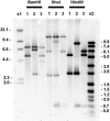
Southern blot of 1 μg of chromosomal DNA from the parental strain and selected integration clones hybridized with an msa ORF probe. Chromosomal DNAs, digested with the indicated enzyme, were from the parental ATCC type strain 33209 (lanes 1), one clone bearing msa1 integrations (lanes 2), and one clone bearing msa2 integrations (lanes 3). Lane s1 contains molecular mass marker II (Boehringer Mannheim), and relative masses are labeled to the left. Lane s2 contains molecular mass marker VII (Boehringer Mannheim), and relative masses are labeled to the right. Sizes are in kilobases. Blot images were captured with an ImageStation 440CF using 1D Image Analysis software, v. 3.5.3 (Kodak).
FIG. 3.
FIG. 3.
Schematic representation of the organization of the chromosomal integration sites at the msa1 locus (A) or msa2 locus (B). Chromosomal sequences are represented by thick lines, and plasmid sequences are represented by thin lines. The plasmid gene conferring resistance to kanamycin is labeled “kan.” The msa ORF fragment in pACP41 is a shaded box. The approximate position of an in-frame stop codon is indicated by a short, vertical arrow. The position of the msa ORF probe used in Southern hybridizations is shown by a diagonally hatched box beneath the msa1 figure. Restriction sites are indicated beneath each figure as follows: B, BamHI; H, HindIII; X, XhoI.
FIG. 4.
FIG. 4.
Western blot of cell-associated extract (CA), cell surface-associated extract (CSE), and supernatant (SUP) from broth cultures of the parental strain (lanes 1), a clone bearing an msa1 disruption (lanes 2), and a clone bearing an msa2 disruption (lanes 3). Cell-equivalent amounts of protein for each strain were analyzed, and MSA protein was identified with monoclonal antibody. Lane m contains molecular mass standards, and indicated sizes are in kilodaltons. The blot image was captured with an ImageStation 440CF using 1D Image Analysis software, v. 3.5.3 (Kodak).
FIG. 5.
FIG. 5.
Cumulative percent survival in groups of chinook salmon challenged by intraperitoneal injection with PBS-peptone (vehicle control) or one of the R. salmoninarum strains. Fish were held in 12°C water and monitored daily for 115 days after challenge; data markers are shown at 10-day intervals. (a) Survival of fish injected with the parental type strain ATCC 33209 or with a clone bearing an msa2 disruption (AMC2). (b) Survival of fish injected with the parental type strain 33209 or with a clone bearing an msa1 disruption (AMC1).

Similar articles

Cited by

References

    1. Alcorn, S., A. L. Murray, R. J. Pascho, and J. Varney. 2005. A cohabitation challenge to compare the efficacies of vaccines for bacterial kidney disease (BKD) in chinook salmon Oncorhynchus tshawytscha. Dis. Aquat. Organ. 63:151-160. - PubMed
    1. Balfry, S. K., L. J. Albright, and T. P. T. Evelyn. 1996. Horizontal transfer of Renibacterium salmoninarum among farmed salmonids via the fecal-oral route. Dis. Aquat. Organ. 25:1-2.
    1. Brown, L. L., G. K. Iwama, and T. P. T. Evelyn. 1996. The effect of early exposure of coho salmon (Oncorhynchus kisutch) eggs to the p57 protein of Renibacterium salmoninarum on the development of immunity to the pathogen. Fish Shellfish Immunol. 6:149-165.
    1. Bruno, D. W. 2004. Prevalence and diagnosis of bacterial kidney disease (BKD) in Scotland between 1990 and 2002. Dis. Aquat. Organ. 59:125-130. - PubMed
    1. Bruno, D. W. 1988. The relationship between auto-agglutination, cell surface hydrophobicity and virulence of the fish pathogen Renibacterium salmoninarum. FEMS Microbiol. Lett. 51:135-140.

Publication types

LinkOut - more resources