

Toxigenic Vibrio cholerae in the aquatic environment of Mathbaria, Bangladesh
- PMID: 16597991
- PMCID: PMC1449004
- DOI: 10.1128/AEM.72.4.2849-2855.2006
Toxigenic Vibrio cholerae in the aquatic environment of Mathbaria, Bangladesh
Abstract
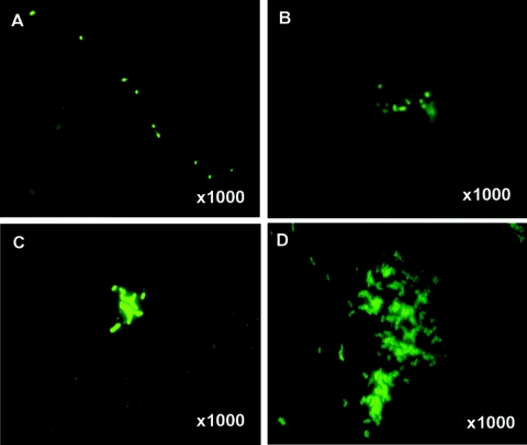
Toxigenic Vibrio cholerae, rarely isolated from the aquatic environment between cholera epidemics, can be detected in what is now understood to be a dormant stage, i.e., viable but nonculturable when standard bacteriological methods are used. In the research reported here, biofilms have proved to be a source of culturable V. cholerae, even in nonepidemic periods. Biweekly environmental surveillance for V. cholerae was carried out in Mathbaria, an area of cholera endemicity adjacent to the Bay of Bengal, with the focus on V. cholerae O1 and O139 Bengal. A total of 297 samples of water, phytoplankton, and zooplankton were collected between March and December 2004, yielding eight V. cholerae O1 and four O139 Bengal isolates. A combination of culture methods, multiplex-PCR, and direct fluorescent antibody (DFA) counting revealed the Mathbaria aquatic environment to be a reservoir for V. cholerae O1 and O139 Bengal. DFA results showed significant clumping of the bacteria during the interepidemic period for cholera, and the fluorescent micrographs revealed large numbers of V. cholerae O1 in thin films of exopolysaccharides (biofilm). A similar clumping of V. cholerae O1 was also observed in samples collected from Matlab, Bangladesh, where cholera also is endemic. Thus, the results of the study provided in situ evidence for V. cholerae O1 and O139 in the aquatic environment, predominantly as viable but nonculturable cells and culturable cells in biofilm consortia. The biofilm community is concluded to be an additional reservoir of cholera bacteria in the aquatic environment between seasonal epidemics of cholera in Bangladesh.
Figures


References
-
- Albert, M. J., A. K. Siddique, M. S. Islam, A. S. G. Faruque, M. Ansaruzzamam, S. M. Faruque, and R. B. Sack. 1993. A large outbreak of clinical cholera due to Vibrio cholerae non-O1 in Bangladesh. Lancet 341:704. - PubMed
-
- Caldwell, D. E. 1995. Cultivation and study of biofilm communities, p. 64-79. In H. M. L. Scott and J. W. Costerton (ed.), Microbial biofilms. University Press, Cambridge, United Kingdom.
-
- Colwell, R. R., P. R. Brayton, D. B. Roszak, A. Huq, and L. M. Palmer. 1985. Viable but non-culturable Vibrio cholerae and related pathogens in the environment: implications for release of genetically engineered microorganisms. Bio/Technology 3:817-820.
-
- Colwell, R. R., and W. M. Spira. 1992. The ecology of Vibrio cholerae, p. 107-127. In D. Barua and W. B. I. Greenouh (ed.), Cholera. Plenum Press, Inc., New York, N.Y.
Publication types
MeSH terms
Substances
Grants and funding
LinkOut - more resources
Full Text Sources
