

doi: 10.1128/AEM.72.4.3016-3020.2006.
Fluorescence in situ hybridization and spectral imaging of coral-associated bacterial communities
Affiliations
- PMID: 16598010
- PMCID: PMC1449077
- DOI: 10.1128/AEM.72.4.3016-3020.2006
Item in Clipboard
Fluorescence in situ hybridization and spectral imaging of coral-associated bacterial communities
Appl Environ Microbiol.
2006 Apr.
Abstract
Microbial communities play important roles in the functioning of coral reef communities. However, extensive autofluorescence of coral tissues and endosymbionts limits the application of standard fluorescence in situ hybridization (FISH) techniques for the identification of the coral-associated bacterial communities. This study overcomes these limitations by combining FISH and spectral imaging.
Figures
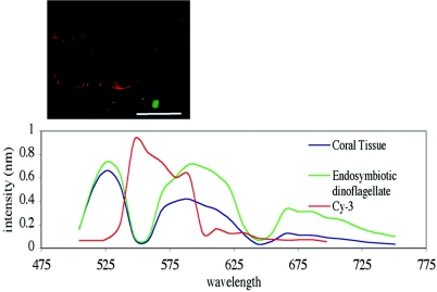
Spectral imaging of A. aspera coral tissue, endosymbiotic dinoflagellate autofluoresence, and EUBmix-Cy3-labeled E. coli. Red, bacteria; blue, coral tissue; green, endosymbiotic dinoflagellates. Scale bar, 50 μm.
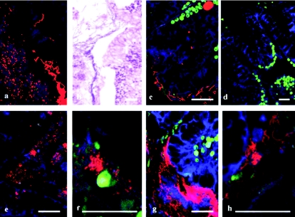
Bacterial communities associated with the lesion created by predation of A. formosa by Drupella sp. FISH identified two bacterial morphologies using EUBmix-Cy3 (a) which were also evident in adjacent sections using hematoxylin and eosin staining (b) and coating the surface of adjacent coral tissues (c). Nonspecific staining of the FISH protocol was not evident (d), and the two bacterial morphologies were identified as a coccoid γ-proteobacterium using the probes GAM42a (e and f) and as a filamentous Cytophaga-Flavobacterium using the probe CF319 (g and h). Red, bacteria; blue, coral tissue; green, endosymbiotic dinoflagellates. Scale bar, 50 μm.
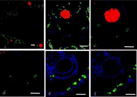
FISH using universal bacterial probe EUBmix of large bacterial aggregates associated with coral tissues S. pistillata and A. formosa (a and b) and identification as γ-proteobacteria using GAM42a (c), with no binding of CF319 (d), HGC69a (e), or LGC 354 (f) probes. Bacterial aggregates, red; coral tissue, blue; endosymbiotic dinoflagellates, green. Scale bar, 50 μm.
References
Publication types
MeSH terms
Substances
LinkOut - more resources
Full Text Sources
Other Literature Sources
