. 2006 Apr 15;20(8):977-89.
doi: 10.1101/gad.1396306. Epub 2006 Apr 5.

The Drosophila P68 RNA helicase regulates transcriptional deactivation by promoting RNA release from chromatin

Affiliations

The Drosophila P68 RNA helicase regulates transcriptional deactivation by promoting RNA release from chromatin

Michael Buszczak et al. Genes Dev. .

Abstract

Terminating a gene's activity requires that pre-existing transcripts be matured or destroyed and that the local chromatin structure be returned to an inactive configuration. Here we show that the Drosophila homolog of the mammalian P68 RNA helicase plays a novel role in RNA export and gene deactivation. p68 mutations phenotypically resemble mutations in small bristles (sbr), the Drosophila homolog of the human mRNA export factor NXF1. Full-length hsp70 mRNA accumulates in the nucleus near its sites of transcription following heat shock of p68 homozygotes, and hsp70 gene shutdown is delayed. Unstressed mutant larvae show similar defects in transcript accumulation and gene repression at diverse loci, and we find that p68 mutations are allelic to Lighten-up, a known suppressor of position effect variegation. Our observations reveal a strong connection between transcript clearance and gene repression. P68 may be needed to rapidly remove transcripts from a gene before its activity can be shut down and its chromatin reset to an inactive state.


Figures

Figure 1.
Figure 1.
Drosophila P68 localizes to specific sites on polytene chromosomes. (A) A CB02119/TM3 third instar larval salivary gland cell in which the EGFP-P68 fusion protein (green) localizes around the rim of the nucleolus (marked by anti-AJ1 staining in red) and with specific bands on polytene chromosomes (DNA marked in blue). (B) p68 gene and transcript structure. The p68 gene (solid line) encodes at least six different normal transcripts (four are shown); the RA transcript fuses to the EGFP exon (green) carried by CB02119. The untranslated region (white), coding regions (gray), and insertion alleles (triangles) are also shown. The red line above the gene structure represents the N-terminal region against which the anti-P68 polyclonal antibody was generated. (C) Wild-type polytene chromosomes stained with anti-P11 (red) and anti-P68 (green) antibodies. Endogenous P68 is indistinguishable from CB02119 EGFP expression, and colocalizes with P11 on chromosomal puffs and other secondary loci. (D) Scanning electron micrographs showing normal dorsocentral and scutellar bristles (arrows) from wild type that are reduced or missing in an CB02119/l(3)01084 adult. (E) Wild-type and CB02119/l(3)01084 stage 5–7 ovarian follicles stained with DAPI. The nurse cell chromatin in p68 mutant follicles frequently fails to disperse as in wild type (cf. arrows in wild-type [WT] and p68−/−samples). (F) Western blot of ovarian and larval extracts probed with anti-P68 antibody. In lanes corresponding to control ovarian and larval samples, the anti-P68 antibody cross-reacts with proteins of the expected molecular weights for the endogenous P68 isoforms (arrowhead). The CB02119 allele expresses a small amount of normal P68. In addition, other bands corresponding to proteins with the mobility of endogenous P68 plus EGFP (bracket) are observed. The l(3)01084, LipF, and LipH alleles are inferred to express very little P68. Anti-ACTIN antibodies were used for the loading control.
Figure 2.
Figure 2.
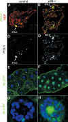
p68 mutants display RNA export defects. (A,B) Control (A) and CB02119/LipF (B) transheterozygotes labeled for hsp70 RNA immediately after a 20-min heat shock. The edge of representative nuclei is outlined in white (boxes). p68 mutant salivary glands have less cytoplasmic hsp70 RNA and qualitatively higher levels of hsp70 RNA at transcription sites. (C,D) Control (C) and CB02119/LipF (D) salivary glands labeled with Br-UTP. Control cells exhibit cytoplasmic staining and punctuate nuclear Br-UTP staining, whereas p68 mutant salivary glands display robust Br-UTP labeling on polytene chromosomal bands. Little signal was observed in the cytoplasm of mutant cells. (E–H) Control (E,G) and CB02119/LipF (F,H) salivary gland cells stained with anti-SBR (NXF1) antibodies (E,F) or anti-ALY (REF1) antibodies (G,H). SBR and ALY are found along the nuclear periphery and in the nucleoplasm of control cells. In p68 mutant cells, their distribution is changed, and both export factors accumulate in the nuclear interior.
Figure 3.
Figure 3.
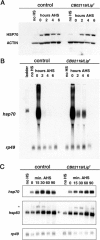
p68 mutants exhibit both delays in translation and prolonged gene transcription. (A) Western blot analysis of larval extracts probed with anti-HSP70 and anti-ACTIN antibodies. No heat shock (no HS) represents non-heat-shocked controls. Animals were heat-shocked for 30 min and allowed to recover for the indicated times after heat shock. HSP70 expression is detectable at the 0-h time point in the control sample, increased up to 2 h AHS, and then begins to decline. In mutant samples, HSP70 expression is undetectable immediately after a 30-min heat shock (0 h AHS), is first detected at the 2-h AHS time point, and continues to increase for up to 6 h AHS. (B) Northern blot analysis of heat-shocked animals. Control and CB02119/LipF mutant animals were heat-shocked for 30 min and allowed to recover for up to 6 h. Controls displayed robust expression of hsp70 in response to heat shock that rapidly declined upon removal from heat shock. CB02119/LipF mutant samples continued to express hsp70 for up to 4 h after removal from heat shock. (C) Control and CB02119/LipF mutant animals were heat-shocked (HS) for 10 min and allowed to recover for the indicated times. Total RNA was isolated, separated on a formaldehyde gel, and probed for hsp70, hsp83, and rp49 RNA. Control and p68 mutant samples do not display any overt differences in the timing or expression levels of hsp70 and hsp83 during the initial response to a short heat shock. Furthermore, the amount of unspliced hsp83 (asterisk) appears to be similar in control and p68 mutant samples, suggesting that loss of p68 does not affect RNA processing.
Figure 4.
Figure 4.
Reduced P68 activity results in persistent transcription factor binding to chromosomes. (A) Control and CB02119/LipF polytene chromosomes from unstressed larvae stained with anti-H3phosS10 and P11 antibodies. (B) Control and CB02119/LipF polytene chromosome squashes from larvae heat-shocked for 20 min and dissected immediately (0 min AHS) or 60 min AHS. These samples were stained with anti-H3phosS10 (green) and anti-HSF (red) antibodies. HSF binding to heat-shock response puffs appears to be comparable in control and mutant samples immediately after heat shock. However, the level and number of sites labeled with the anti-H3phosS10 antibody are greater in p68 mutants. While HSF is lost from heat-shock puffs in control samples (control inset) 60 min AHS, HSF binding persists in p68 mutant chromosomes (p68−/− inset). Insets show the hsp70 loci at 87A and 87C.
Figure 5.
Figure 5.
p68 mutation blocks gene shutoff during heat shock. (A–D) Control (A,C) and CB02119/LipH (B,D) larvae were heat-shocked at 37°C for 20 min and immediately processed. The cells were stained using anti-H5 antibodies (green) and anti-HSF antibodies (red) (cf. C and D). Examining the anti-H5 staining alone reveals that active Pol II is limited to major heat-shock puffs in control cells (C), while in p68 mutant cells, it is present at heat-shock loci, other discrete sites, and generally along chromosome arms (D) (hsp70 gene loci at 87A and 87C are indicated). (E–F) Control (E,G) and CB02119/LipH (F,H) larvae were heat-shocked at 37°C for 10 min, pulse-labeled with Br-UTP, and heat-shocked for an additional 20 min. (E,G) Control samples exhibit Br-UTP labeling only at heat-shock response puffs. (F,H) p68 mutant cells show high levels of Br-UTP incorporation at many chromosomal sites including the nucleolus, demonstrating that a broad number of genes continue to transcribe RNA during heat shock in the absence of p68.
Figure 6.
Figure 6.
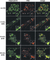
The localization of P68 changes during heat shock. Salivary gland polytene chromosome squashes from wild-type wandering third instar larvae. Samples were unstressed (no HS) or subjected to a 30-min heat shock at 37°C followed by no recovery period (0 min AHS), 10 min of recovery at room temperature (10 min AHS), and 30 min of recovery (30 min AHS). Squashes were stained with anti-H3phosS10 and anti-P68 antibodies. Relevant cytological loci are indicated in each panel. In non-heat-shocked animals, there is robust colocalization between H3phosS10 and P68 on a few developmental puffs. Upon heat shock, H3phosS10 accumulates at heat-shock puffs (87A,C) and decreases at other sites (74E,F; 75B). However, P68 remains bound to developmental puffs. After a 10-min recovery period, P68 begins to colocalize with H3phosS10 on heat-shock puffs. This accumulation of P68 on heat-shock puffs continues to increase up to 30 min after heat shock. Thirty minutes into recovery, H3phosS10 staining begins to accumulate on other bands, while P68 remains highly enriched on heat-shock puffs.
Figure 7.
Figure 7.
Ecdysone response genes exhibit prolonged expression in the absence of p68. (A) RT–PCR reactions using E74A, E74B, and rp49 specific primers. Total RNA was isolated from control and CB02119/LipF animals at the various points after puparium formation. Expression of E74B in controls peaks at −3 h APF and then decreases from 2 to 10 h APF. A second peak of E74B expression is observed beginning at 12 h APF. E74A expression in control animals first peaks at 2 h APF and then declines to low levels between 6 and 8 h APF, followed by a second peak at 10 h APF that declines rapidly. Mutant animals exhibit prolonged expression of both E74A and E74B between 2 and 8 h APF. In addition, the second peak of E74A expression never declines in mutant animals over the time course of the experiment. (B) RT–PCR using E75A- and E75B-specific primers. p68 mutant animals exhibit prolonged expression of both E75A and E75B relative to the control samples.

References

    1. Adelman K., Marr M.T., Werner J., Saunders A., Ni Z., Andrulis E.D., Lis J.T. Efficient release from promoter-proximal stall sites requires transcript cleavage factor TFIIS. Mol. Cell. 2005;17:103–112. - PubMed
    1. Aguilera A. Cotranscriptional mRNP assembly: From the DNA to the nuclear pore. Curr. Opin. Cell Biol. 2005;17:242–250. - PubMed
    1. Ali A., Bharadwaj S., O’Carroll R., Ovsenek N. HSP90 interacts with and regulates the activity of heat shock factor 1 in Xenopus oocytes. Mol. Cell. Biol. 1998;18:4949–4960. - PMC - PubMed
    1. Andrulis E.D., Guzman E., Doring P., Werner J., Lis J.T. High-resolution localization of Drosophila Spt5 and Spt6 at heat shock genes in vivo: Roles in promoter proximal pausing and transcription elongation. Genes & Dev. 2000;14:2635–2649. - PMC - PubMed
    1. Andrulis E.D., Werner J., Nazarian A., Erdjument-Bromage H., Tempst P., Lis J.T. The RNA processing exosome is linked to elongating RNA polymerase II in Drosophila. Nature. 2002;420:837–841. - PubMed

Publication types

MeSH terms

LinkOut - more resources