. 2006 Apr 15;20(8):966-76.
doi: 10.1101/gad.1404206. Epub 2006 Apr 5.

Histone sumoylation is a negative regulator in Saccharomyces cerevisiae and shows dynamic interplay with positive-acting histone modifications

Affiliations

Histone sumoylation is a negative regulator in Saccharomyces cerevisiae and shows dynamic interplay with positive-acting histone modifications

Dafna Nathan et al. Genes Dev. .

Abstract

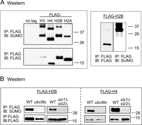
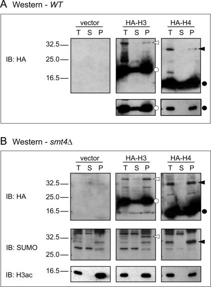
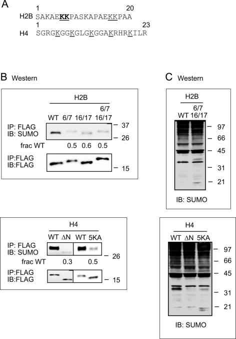
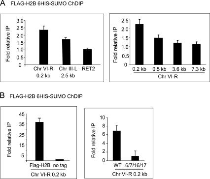
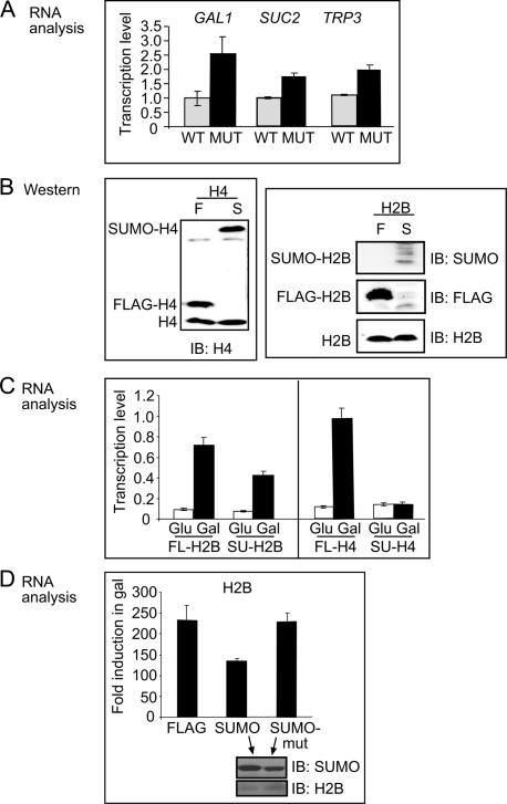
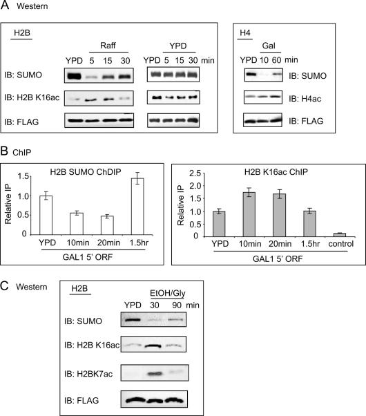
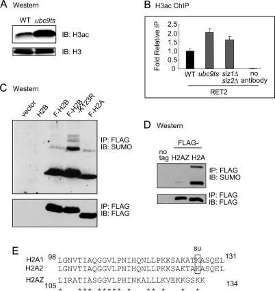
Covalent histone post-translational modifications such as acetylation, methylation, phosphorylation, and ubiquitylation play pivotal roles in regulating many cellular processes, including transcription, response to DNA damage, and epigenetic control. Although positive-acting post-translational modifications have been studied in Saccharomyces cerevisiae, histone modifications that are associated with transcriptional repression have not been shown to occur in this yeast. Here, we provide evidence that histone sumoylation negatively regulates transcription in S. cerevisiae. We show that all four core histones are sumoylated and identify specific sites of sumoylation in histones H2A, H2B, and H4. We demonstrate that histone sumoylation sites are involved directly in transcriptional repression. Further, while histone sumoylation occurs at all loci tested throughout the genome, slightly higher levels occur proximal to telomeres. We observe a dynamic interplay between histone sumoylation and either acetylation or ubiquitylation, where sumoylation serves as a potential block to these activating modifications. These results indicate that sumoylation is the first negative histone modification to be identified in S. cerevisiae and further suggest that sumoylation may serve as a general dynamic mark to oppose transcription.


Figures

Figure 1.
Figure 1.
In vivo histone sumoylation in yeast. (A) Western blot (IB) analysis of immunoprecipitated (IP) Flag-tagged histones using anti-Flag and anti-SUMO antibodies. Flag-tagged histones migrate ∼15 kDa. Singly sumoylated histones migrate between 26 and 37 kDa. (B) Flag IP followed by Western blot (IB) analysis of histones in sumoylation pathway mutant strains ubc9ts and siz1Δsiz2Δ. Detection was carried out as in A.
Figure 2.
Figure 2.
Western blot analysis of histones in fractionated yeast cells. (A) Western blot analysis using anti-HA antibody of untagged (vector) fractionated wild-type cells or HA-tagged H3 or H4. Fractions are total (T), soluble (S), or pellet (P). Open circles indicate HA-H3, closed circles indicate HA-H4, open arrowheads point to sumoylated HA-H3, and closed arrowheads point to sumoylated HA-H4. A shorter exposure of the nonsumoylated histones is shown below. (B) Similar analysis as in A except extracts were obtained from a strain bearing a SMT4 (SUMO protease) deletion, which increases the cellular level of sumoylated histones. HA and SUMO immunoblots are shown, as well as H3 acetylation (H3ac) immunoblot as a control for fractionation.
Figure 3.
Figure 3.
Identification and confirmation of histone SUMO sites in vivo. (A) Regions of sumoylation in histone H2B and H4. The lysine residues for which sumoylation was confirmed by MS/MS in histone H2B are shown in bold and underlined letters. Additional putative sumoylation sites in H2B are also underlined. Lysine residues that were mutated to alanine in histone H4 are indicated by underlined letters. (B) Effect of substitution mutations in putative sumoylation sites on in vivo modification in yeast. Lys6 and Lys7 (6/7), Lys16 and Lys17 (16/17), or all four lysines (6/7 16/17) in H2B were mutated to alanine and were either shuffled in as the only source of histone H2B in the cell or transformed and kept as an ectopic copy. Histone H4 was expressed ectopically, and the entire tail was deleted (ΔN), or all five lysines in the H4 tail were mutated to alanine (5K). Sumoylation levels were detected using Flag IP followed by Western blot analysis with anti-Flag (bottom) and anti-SUMO (top) antibodies. The numbers below the SUMO blots represent quantification of the sumoylation levels compared with the signal in wild-type (WT) strain after normalization to the Flag levels in each strain. (C) Whole-cell extracts from the same strains used in B were analyzed by SDS-PAGE and immunoblotting with an anti-SUMO antibody. At higher molecular weight (above the 97-kDa marker), the lanes are smeared and distinct bands are no longer seen. The H4 wild-type (WT) lane shows a band ∼33 kDa, which corresponds to the molecular weight of sumoylated H4, and is greatly reduced in the H4 mutant lanes. The H2B genes were not on a high-copy plasmid as were the H4 genes; therefore, the sumoylated form of H2B is not visible in whole-cell extracts.
Figure 4.
Figure 4.
Location of histone sumoylation in the yeast genome. (A) Quantitative PCR analysis of ChDIP in a His-SUMO Flag-H2B strain. Telomeric end primers are located ∼0.2 kb away from the telomeric repeats on Chr VI-R or 2.5 kb away from telomeric repeats of Chr III-L. The right panel shows ChDIP using primers to regions 0.5, 3.6, and 7.3 kb away from the telomere end on Chr VI-R. RET2 is located ∼20 kb away from the Chr VI-R telomere end. (B) Controls for quantitative PCR analysis of ChDIP samples. All strains have His-tagged SUMO. Left panel shows a comparison control stain lacking the Flag-H2B plasmid. Right panel shows a comparison control strain containing H2B substitution mutant K6/7/16/17A in the Flag-H2B plasmid.
Figure 5.
Figure 5.
Sumoylation of histones regulates transcription. (A) Quantitative PCR analysis of TRP3, SUC2, and GAL1 RNA levels in noninducing (YPD) conditions for wild-type (WT) and H2B K6/7/16/17A substitution mutant (MUT). (B) Western blot analysis of whole-cell extracts probed with anti-histone H4 (left) or anti-histone H2B, anti-Flag, and anti-SUMO antibodies (panels on right). Indicated are the positions of endogenous (bottom), Flag-tagged histones (middle), and SUMO-fused histones (top). (C) Quantitative PCR analysis showing transcript levels of GAL1 for SUMO–H2B (SU-H2B) or Flag-H2B (FL-H2B) fusions when switching from glucose (Glu) to galactose (Gal) media and the parallels in histone H4. (D) Quantitative PCR analysis as in C comparing Flag-H2B and SUMO–H2B with a strain with a SUMO–H2B fusion containing substitution mutations in SUMO at Lys37 and Arg46 to glutamic acid (SUMO-mut). Lower insert shows Western analysis of whole-cell extracts from SUMO–H2B and SUMO-mut–H2B fusion strains using anti-SUMO and anti-H2B antibodies.
Figure 6.
Figure 6.
Interplay of acetylation–sumoylation in histone H2B and H4 upon changes in carbon source. (A) Flag IP followed by Western blot analysis of Flag-H2B (left and middle) or Flag-H4 (right) samples taken at the indicated time points during growth switched from glucose-to-raffinose (Raff, H2B) or glucose-to-galactose (Gal, H4). Center panels show the control Glu-to-Glu switch for Flag H2B. Antibodies are as indicated. (B) Real-time PCR analysis at the GAL1 promoter region of a ChDIP experiment (left) or H2B K16ac ChIP (right) in a His-SUMO Flag-H2B tagged strain. Samples were taken at the indicated time points after switching from glucose (YPD) to raffinose. (C) Flag IP followed by Western blot analysis of Flag-H2B samples taken at the indicated time points during glucose (YPD) to ethanol/glycerol (EtOH/Gly) carbon source change. Antibodies are as indicated.
Figure 7.
Figure 7.
Relationship between sumoylation and postive histone mechanisms. (A) Western blot of histones prepared from wild-type (WT) or ubc9ts strain probed with H3ac (K9ac, K14ac) antibody (top) or unmodified histone H3 antibody (bottom). (B) Quantitative PCR analysis of H3ac ChIP of the RET2 gene in the wild-type (WT), ubc9ts, and siz1Δsiz2Δ strains. (C) Western blot of histones prepared from wild-type (WT) strain or strains bearing Flag-H2B, Flag-H2B-K123R substitution mutation, or Flag-H2A. (D) Flag IP followed by Western blot analysis of Flag-tagged histone H2A and histone variant H2AZ. (E) Alignment of the C-terminal tails of yeast canonical H2A and variant histone H2AZ. The sumoylation site is indicated for H2A. Asterisks indicate identical residues.

References

    1. Aparicio O.M., Billington B.L., Gottschling D.E. Modifiers of position effect are shared between telomeric and silent mating-type loci in S. cerevisiae. Cell. 1991;66:1279–1287. - PubMed
    1. Bannister A.J., Zegerman P., Partridge J.F., Miska E.A., Thomas J.O., Allshire R.C., Kouzarides T. Selective recognition of methylated lysine 9 on histone H3 by the HP1 chromo domain. Nature. 2001;410:120–124. - PubMed
    1. Berger S.L. Histone modifications in transcriptional regulation. Curr. Opin. Genet. Dev. 2002;12:142–148. - PubMed
    1. Biggins S., Bhalla N., Chang A., Smith D.L., Murray A.W. Genes involved in sister chromatid separation and segregation in the budding yeast Saccharomyces cerevisiae. Genetics. 2001;159:453–470. - PMC - PubMed
    1. Boudreault A.A., Cronier D., Selleck W., Lacoste N., Utley R.T., Allard S., Savard J., Lane W.S., Tan S., Cote J. Yeast enhancer of polycomb defines global Esa1-dependent acetylation of chromatin. Genes & Dev. 2003;17:1415–1428. - PMC - PubMed

Publication types