. 2006 Apr 15;20(8):954-65.
doi: 10.1101/gad.1409106. Epub 2006 Apr 5.

The role of Rat1 in coupling mRNA 3'-end processing to transcription termination: implications for a unified allosteric-torpedo model

Affiliations

The role of Rat1 in coupling mRNA 3'-end processing to transcription termination: implications for a unified allosteric-torpedo model

Weifei Luo et al. Genes Dev. .

Abstract

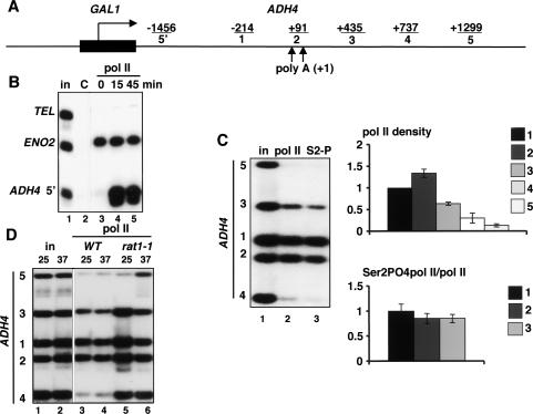
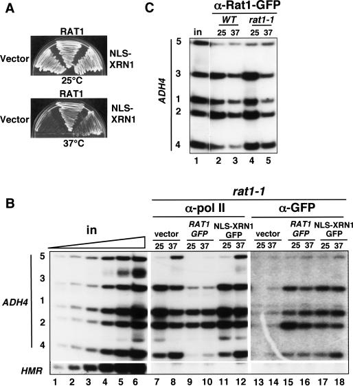
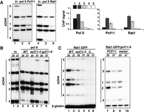
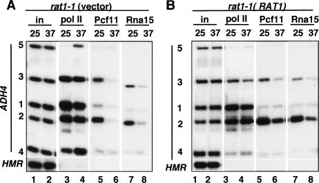
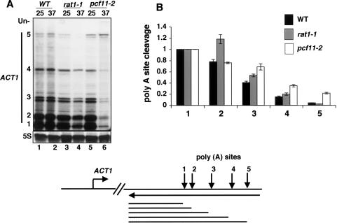
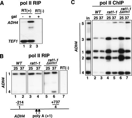
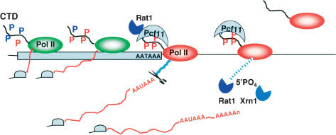
The torpedo model of transcription termination by RNA polymerase II proposes that a 5'-3' RNA exonuclease enters at the poly(A) cleavage site, degrades the nascent RNA, and eventually displaces polymerase from the DNA. Cotranscriptional degradation of nascent RNA has not been directly demonstrated, however. Here we report that two exonucleases, Rat1 and Xrn1, both contribute to cotranscriptional degradation of nascent RNA, but this degradation is not sufficient to cause polymerase release. Unexpectedly, Rat1 functions in both 3'-end processing and termination by enhancing recruitment of 3'-end processing factors, including Pcf11 and Rna15. In addition, the cleavage factor Pcf11 reciprocally aids in recruitment of Rat1 to the elongation complex. Our results suggest a unified allosteric/torpedo model in which Rat1 is not a dedicated termination factor, but is an integrated component of the cleavage/polyadenylation apparatus.


Figures

Figure 1.
Figure 1.
Transcription termination at the chromosomal ADH4 gene. (A) Diagram of GAL1-ADH4. PCR products are indicated with the distances in base pairs from the center of each to the major poly(A) site (+1). Upward arrows indicate the two poly(A) sites. (B) Inducible transcription of GAL1-ADH4. Pol II ChIP of GAL1-ADH4 in wild-type cells (DBY548) induced with galactose for the indicated times. The ADH4 5′ primer pair (−1456 in A) was used. ENO2 is a positive control, and telomere VIR (TEL) is a nontranscribed negative control. IPs were with polyclonal anti-pan CTD that detects total pol II. (in) Input chromatin; (C) control IP with irrelevant antibody. (C) Transcription termination and CTD Ser2 phosphorylation. Anti-pol II ChIP with anti-pan CTD as in B, and anti-Ser2 phosphorylated CTD (S2) in the wild-type strain DBY548. Total pol II density relative to the value at position 1 in the ORF, normalized to input, is shown in the top graph. Ser2 phosphorylated pol II, normalized to total pol II, is shown in the bottom graph. (D) Termination at ADH4 is inhibited by inactivation of Rat1. Pol II ChIP of wild-type (DBY548, lanes 3,4) and rat1-1 (DBY745, lanes 5,6). Note the delayed termination with high pol II at positions 3 and 4 in rat1-1 at 25°C (lane 5) relative to wild type (lane 3).
Figure 2.
Figure 2.
Nuclear Xrn1 does not rescue the termination defect in rat1-1. (A) NLS-Xrn1 complements the ts growth of rat1-1. The rat1-1 strain (DBY745) was transformed with control vector plasmid (pFL38), WTRat1-GFP (pAJ226), or NLS-Xrn1-GFP (pAJ237) and grown at 25°C or 37°C on SC-Ura plates. As shown previously, NLS-Xrn1 complements growth at 37°C (Johnson 1997). (B) NLS-Xrn1 is recruited to the gene, but it does not complement the termination defect in rat1-1. Anti-pol II (lanes 7–12) and anti-GFP (lanes 13–18) ChIP of galactose-induced GAL1-ADH4 in rat1-1 transformed with vector, RAT1-GFP, or NLS-XRN1-GFP plasmids (DBY768, DBY754, and DBY777). (Lanes 1–6) Titration of input demonstrates linearity of the PCR. HMR is a transcriptionally silent control that was run on a separate gel. Note the failure to terminate at 37°C with NLS-Xrn1 (lane 12) and the cross-linking of NLS-Xrn1 to the ADH4 gene (lanes 17,18). (C) Wild-type and ts mutant Rat1 proteins are recruited to the 3′ end at 25°C and 37°C. ChIP of GFP-tagged Rat1 and Rat1-1 on galactose-induced GAL1-ADH4 in DBY786 (WT) and DBY787 (rat1-1).
Figure 3.
Figure 3.
Coordinate recruitment of Rat1 and the 3′-processing factor Pcf11. (A) Highly coincident patterns of Pcf11 and Rat1 recruitment at the ADH4 3′ end. (Left) Pol II, Pcf11, and Rat1-GFP ChIP on galactose-induced GAL1-ADH4 in wild-type cells (DBY786) at 25°C. (Right) ChIP signals normalized to input were normalized relative to the value at position 1 in the ORF. Means and standard deviations from three experiments are shown in the graphs. (B) Termination defects in pcf11-2 and pcf11-9 mutants. Pol II ChIP of wild type (WT), pcf11-2, and pcf11-9 (DBY548, DBY585, DBY593) at 25°C and 37°C. Note delayed termination in pcf11-2 at 25°C (lane 5) and failure to terminate at 37°C (lane 6). (C) Rat1 recruitment to the ADH4 3′ end requires functional Pcf11. (Lanes 1–6) Rat1-GFP ChIP in wild-type (WT) and pcf11-2 strains (DBY786, DBY815) and the untagged control (C, DBY548). (Lanes 7–10) Rat1-GFP ChIP in pcf11-9 (DBY794) transformed with PCF11 plasmid (DBY796) or control vector (DBY795). β-globin is a loading control.
Figure 4.
Figure 4.
Normal recruitment of 3′-processing factors requires functional Rat1. (A) ChIP of pol II, Pcf11, and Rna15 on GAL1-ADH4 in rat1-1 transformed with control vector (DBY 768). (Lanes 6,8) Note recruitment of Pcf11 and Rna15 is reduced by inactivation of Rat1 at 37°C. (B) ChIP as in A of rat1-1 transformed with the wild-type RAT1 plasmid (DBY754). (Lanes 6,8) Note recruitment of Pcf11 and Rna15 at 37°C is partially restored relative to A. HMR-negative controls were run on separate gels.
Figure 5.
Figure 5.
Rat1 is necessary for normal ACT1 poly(A) site selection. (A) ACT1 poly(A) site choice is shifted in favor of more 3′ sites when Rat1 is inactivated. Total RNA from wild-type (WT), rat1-1, or pcf11-2 strains (DBY548, DBY745, and DBY593) grown at 25°C or 37°C for 90 min was analyzed by RPA with 3′ ACT1 and 5S rRNA probes. (Un) Uncleaved RNA. The diagram shows poly(A) cleavage sites 1–5, the ACT1 anti-sense riboprobe, and RNase protection products. (B) Quantification of ACT1 mRNA cleavage at sites 1–5 in wild-type (WT), rat1-1, and pcf11-2 cells at 37°C. Cleavage relative to site 1 is shown after normalization for the 32P-U content of the protection products 1–5. Values are means and standard deviations from two experiments.
Figure 6.
Figure 6.
Degradation of nascent RNA by Rat1 and Xrn1 is not sufficient to cause pol II termination. (A) Anti-pol II RIP detects nascent transcripts. The wild-type strain (DBY548) was analyzed without (−) and with (+) galactose (gal) induction. Immunoprecipitated RNA was analyzed by RT–PCR with primers for GAL1-ADH4 (position 1) and constitutively expressed TEF1. RT(+) and RT(−) signify plus and minus RT, respectively. (B) Nascent RNA is degraded downstream of the poly(A) site. Anti-pol II RIP of GAL1-ADH4 in galactose-induced wild-type (WT), rat1-1, and rat1-1Δxrn1 strains (DBY548, DBY745, and DBY772) at 25°C or 37°C. Lane 7 is a control − RT. Note the absence of nascent RNA associated with pol II that has failed to terminate at position 4 in lanes 3–5 and stabilization of nascent RNA when both Rat1 and Xrn1 were eliminated (lane 6). This result is representative of four independent experiments. PCR product 4 has 65% as many 32P-dC residues as product 1. (C) Anti-pol II ChIP of the samples in B. (Lanes 4–6) Note pol II that failed to terminate is present at high density at position 4, although nascent RNA was not detectable in B.
Figure 7.
Figure 7.
Hybrid model for coupling termination with 3′-end processing. This model incorporates aspects of both the antiterminator/allosteric and torpedo models. Cooperative association of Rat1 and cleavage/polyadenylation factors exemplified by Pcf11 with the CTD Ser2 phosphorylated pol II elongation complex is shown at the poly(A) site (AATAAA). This interaction is proposed to result in an allosteric change in pol II (designated by a change from green to red) that favors termination. (Blue Ps) Ser5-PO4; (red Ps) Ser2-PO4. Nascent RNA downstream of the poly(A) cleavage site (blue line) is degraded by both Xrn1 and Rat1. This degradation could facilitate termination; however, it is not sufficient to cause termination.

Comment in

References

    1. Ahn S.H., Kim M., Buratowski S. Phosphorylation of serine 2 within the RNA polymerase II C-terminal domain couples transcription and 3′ end processing. Mol. Cell. 2004;13:67–76. - PubMed
    1. Amberg D.C., Goldstein A.L., Cole C.N. Isolation and characterization of RAT1: An essential gene of Saccharomyces cerevisiae required for the efficient nucleocytoplasmic trafficking of mRNA. Genes & Dev. 1992;6:1173–1189. - PubMed
    1. Amrani N., Minet M., Wyers F., Dufour M.E., Aggerbeck L.P., Lacroute F. Pcf11 encodes a 3rd protein-component of yeast cleavage and polyadenylation factor I. Mol. Cell. Biol. 1997;17:1102–1109. - PMC - PubMed
    1. Bauren G., Belikov S., Wieslander L. Transcriptional termination in the Balbiani ring 1 gene is closely coupled to 3′-end formation and excision of the 3′-terminal intron. Genes & Dev. 1998;12:2759–2769. - PMC - PubMed
    1. Birse C.E., Lee B.A., Hansen K., Proudfoot N.J. Transcriptional termination signals for RNA polymerase II in fission yeast. EMBO J. 1997;16:3633–3643. - PMC - PubMed

Publication types