. 2007 Jan;44(4):311-21.
doi: 10.1016/j.molimm.2006.02.029. Epub 2006 Apr 4.

ZXDC, a novel zinc finger protein that binds CIITA and activates MHC gene transcription

Affiliations

ZXDC, a novel zinc finger protein that binds CIITA and activates MHC gene transcription

Wafa Al-Kandari et al. Mol Immunol. 2007 Jan.

Abstract

The class II trans-activator (CIITA) is recognized as the master regulator of major histocompatibility complex (MHC) class II gene transcription and contributes to the transcription of MHC class I genes. To better understand the function of CIITA, we performed yeast two-hybrid with the C-terminal 807 amino acids of CIITA, and cloned a novel human cDNA named zinc finger, X-linked, duplicated family member C (ZXDC). The 858 amino acid ZXDC protein contains 10 zinc fingers and a transcriptional activation domain, and was found to interact with the region of CIITA containing leucine-rich repeats. Over-expression of ZXDC in human cell lines resulted in super-activation of MHC class I and class II promoters by CIITA. Conversely, silencing of ZXDC expression reduced the ability of CIITA to activate transcription of MHC class II genes. Given the specific interaction between the ZXDC and CIITA proteins, as well as the effect of ZXDC on MHC gene transcription, it appears that ZXDC is an important regulator of both MHC class I and class II transcription.


Figures

Fig. 1
Fig. 1
Schematic diagram of the 858 amino acid ZXDC protein. Open boxes, zinc fingers; black box, transcriptional activation domain (TAD); gray box, CIITA-binding domain. Region of amino acid homology with ZXDA and ZXDB (73% amino acid identity over approximately 500 amino acids) is indicated by dashed line. The amino acid sequences of the 10 zinc fingers in ZXDC are shown.
Fig. 2
Fig. 2
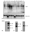
Expression of the ZXDC gene and cDNA. (A) Northern blot of poly-A+ mRNA, probed ZXDC cDNA exons 8–10, outside of the homology region with ZXDA and ZXDB (top panel) or with beta-actin (bottom panel). B, brain; C, colon; H, heart; K, kidney; Li, liver; Lu, lung; M, skeletal muscle; Pl, placenta; SI, small intestine; Sp, spleen; St, stomach; T, testis. (B) Western blot of HEK293 cell extracts from untransfected cells, or cells transfected or with pCMV-ZXDC. Blot was probed with affinity purified anti-ZXDC antiserum. Numerals represent molecular mass of bands, as determined by comparison with molecular mass standards. (C) Western blot of HEK293 cell extracts from untransfected cells with anti-ZXDCantiserum; longer exposure than in panel (B). (D) Western blot with anti-ZXDC antiserum of in vitro transcribed/translated ZXDC protein.
Fig. 3
Fig. 3
ZXDC cooperates with CIITA in activating MHC class I and class II transcription. (A) HEK293 cells were co-transfected with pCMV-CIITA and/or pCMV-ZXDC, along with the reporter plasmid pDRA-luc. pDRA-luc contains the promoter from the HLA-DRA gene (−150/+36) linked to the firefly luciferase gene. Numerals indicate nanograms of each plasmid DNA transfected (total plasmid DNA was kept constant). (B) Similar to (A) except RJ2.2.5 cells were transfected. Numerals indicate micrograms of each plasmid DNA transfected. (C) RJ2.2.5 cells were transfected with pCMV-CIITA and/or pCMV-ZXDC, and HLA-DRA gene transcription measured by quantitative real-time reverse transcriptase PCR. Numerals indicate micrograms of each plasmid DNA transfected. Data were normalized to beta-actin. (D) Transfection was similar to (A) except that the reporter plasmid was pHLAB-luc, which contained the promoter from the HLA-B1 gene (−284/+1) linked to the firefly luciferase gene. Statistically significant differences in activation (by Student’s t-test) are indicated. In all panels, error bars represent standard error of the mean of three independent experiments.
Fig. 4
Fig. 4
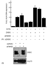
Silencing of ZXDC gene expression results in a reduction in MHC class II activation by CIITA. HEK293 cells were transfected with 1 mg of siRNA directed against ZXDC (or an siRNA with random sequence) followed by transfection with the indicated expression plasmids for ZXDC (700 ng) and/or CIITA (100 ng). (A) HLA-DRA gene expression, 24 h following transfection with plasmid DNA, measured by quantitative real-time RT-PCR. Error bars represent standard error of the mean of three independent experiments. (B) Western analysis with anti-ZXDC antiserum on protein lysates from HEK293 cells transfected with the indicated siRNA. Top panel was probed with anti-ZXDC and bottom panel was probed with anti-Hsp70 as a loading control.
Fig. 5
Fig. 5
Association between CIITA and ZXDC. (A) Co-immunoprecipitation of ZXDC and CIITA. HEK293 cells were transfected with pCMV-CIITA (which has an N-terminal HA-tag) or not transfected, and subjected to immunoprecipitation with anti-ZXDC (or pre-immune serum; data not presented). Western analysis was performed with anti-HA on immunoprecipitates (left panel) or lysates (right panel) to detect the presence of CIITA. (B) Western analysis was performed with anti-ZXDC on immunoprecipitates (left panel) or lysates (right panel) to detect the presence of ZXDC. (C) Chromatin immunoprecipitation with anti-ZXDC, anti-RNA polymerase II, anti-NFYA or non-specific antiserum. Co-immunoprecipitated HLA-DRA promoter DNA (filled bars) or GAPDH promoter DNA (open bars) was quantitated by real-time PCR. Error bars represent the standard error of the mean for three independent experiments.
Fig. 6
Fig. 6
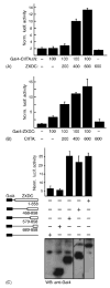
Association of CIITA and ZXDC proteins. (A) Demonstration of ZXDC–CIITA association by mammalian two-hybrid assay. Gal4–CIITAΔN is a fusion between the Gal4 DNA-binding domain and amino acids 323–1130 of CIITA, which lacks the transcriptional activation domain of CIITA. Gal4–CIITAΔN was co-transfected with pG5-luc and increasing amounts of pCMV-ZXDC into HEK293 cells. (B) Gal4–ZXDC is a fusion of the Gal4 DNA-binding domain and the full length ZXDC cDNA. Gal4–ZXDC was co-transfected with pG5-luc and increasing amounts of pCMV-CIITA into HEK293 cells. Note that Gal4–ZXDC activates transcription in the absence of co-expressed CIITA, indicating the presence of a transcriptional activation domain in ZXDC. (C) The transcriptional activation domain of ZXDC was localized by generating serial N-terminal truncations of ZXDC, and expressing them as fusion proteins with the Gal4 DNA-binding domain. The amino acids of ZXDC present in each fusion protein are indicated below the plasmid diagrams. The expression of the Gal4–ZXDC fusion proteins was confirmed by Western blot probed with anti-Gal4. The reporter plasmid for panels (B and C) was pG5-luc, containing five UASg sites linked to the firefly luciferase gene. In panels (B and C), error bars represent standard error of the mean of three independent experiments.
Fig. 7
Fig. 7
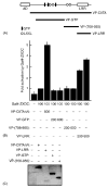
ZXDC binds to the region of CIITA containing leucine-rich repeats. (A) Schematic diagram of CIITA protein indicating conserved domains. AD, transcriptional activation domain; Walker-type GTP-binding motifs (black rectangles); LXXL motifs (open circles); LRR, leucine-rich repeats. Three fragments of the CIITA protein were expressed as fusion proteins with the transcriptional activation domain of herpesvirus VP16. Regions of CIITA protein fused to VP16 activation domain are indicated (see below). Diagram based upon Boss and Jensen (2003). (B) Mammalian two-hybrid transfection experiments in HEK293 cells to determine the ZXDC-binding region of CIITA. The VP-CIITAΔN fusion protein contains amino acids 323–1130 of CIITA; VP-GTP consists of amino acids 336–702 of CIITA, which encompasses the GTP-binding motifs of CIITA; VP-(708–955) consists of amino acids 708–955 of CIITA, which lies between the GTP-binding domain and the leucine-rich repeats (LRR); VP-LRR consists of amino acids 956–1130 of CIITA, which contains the LRR. The reporter plasmid was pG5-luc, containing five UASg sites linked to the firefly luciferase gene. Numerals indicate nanograms of each plasmid DNA transfected (total plasmid DNA was kept constant). The data are representative of four independent experiments. (C) Western analysis of transfected cells with anti-VP16 antiserum, to demonstrate expression of the VP16–CIITA fusion proteins.
Fig. 8
Fig. 8
CIITA binds to the C-terminal 170 amino acids of ZXDC. (A) N-terminal and (B) C-terminal truncations of ZXDC were created as Gal4 fusion proteins. Plasmids expressing the Gal4–ZXDC fusion proteins (100 ng) were co-transfected with, or without, pCMV-CIITA (600 ng) and the reporter plasmid pG5-luc (100 ng) in HEK293 cells. The amino acids of ZXDC present in each fusion protein are indicated below the plasmid diagrams. In both panels, error bars represent standard error of the mean of three independent experiments.
Fig. 9
Fig. 9
The zinc fingers of ZXDC are necessary for full cooperation between CIITA and ZXDC. Plasmids expressing the indicated fragments of ZXDC protein were co-transfected with, or without, pCMV-CIITA and the reporter plasmid pDRA-luc. On the ZXDC protein diagram, the black rectangle represents the 10 zinc fingers (ZnF), the hatched rectangle represents the transcriptional activation domain (AD) and the gray rectangle represents the CIITA-binding domain (CB). Error bars represent standard error of the mean of three independent experiments.

References

    1. Accolla RS. Human B cell variants immunoselected against a single Ia antigen subset have lost expression of several Ia antigen subsets. J Exp Med. 1983;157:1053–1058. - PMC - PubMed
    1. Boss JM, Jensen PE. Transcriptional regulation of the MHC class II antigen presentation pathway. Curr Opin Immunol. 2003;15:105–111. - PubMed
    1. Brown JA, Rogers EM, Boss JM. The MHC class II transactivator (CIITA) requires conserved leucine charged domains for interactions with the conserved W box promoter element. Nucleic Acids Res. 1998;26:4128–4136. - PMC - PubMed
    1. Camacho-Carvajal MM, Klingler S, Schnappauf F, Hake SB, Steimle V. Importance of class II transactivator leucine-rich repeats for dominant-negative function and nucleo-cytoplasmic transport. Int Immunol. 2004;16:65–75. - PubMed
    1. Cress WD, Triezenberg SJ. Critical structural elements of the VP16 transcriptional activation domain. Science. 1991;251:87–90. - PubMed

Publication types

MeSH terms