. 2006 May 15;15(10):1629-39.
doi: 10.1093/hmg/ddl084. Epub 2006 Apr 6.

Tissue-specific roles of Tbx1 in the development of the outer, middle and inner ear, defective in 22q11DS patients

Affiliations

Tissue-specific roles of Tbx1 in the development of the outer, middle and inner ear, defective in 22q11DS patients

Jelena S Arnold et al. Hum Mol Genet. .

Abstract

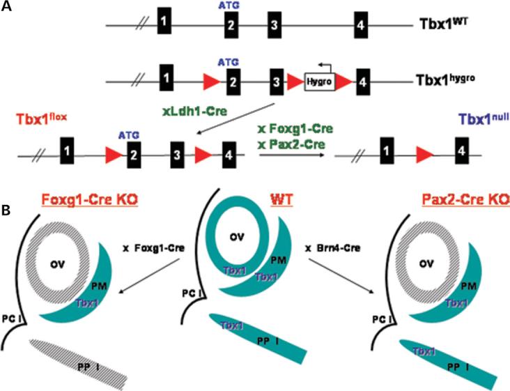
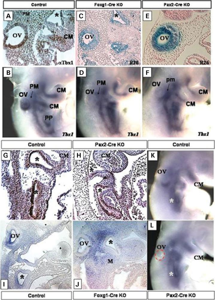
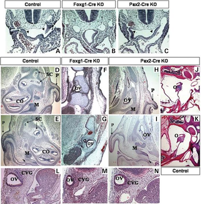
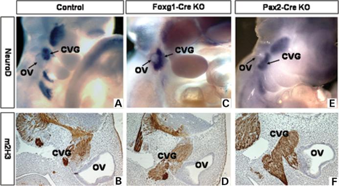
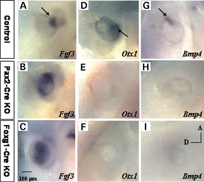
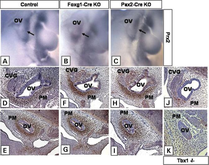
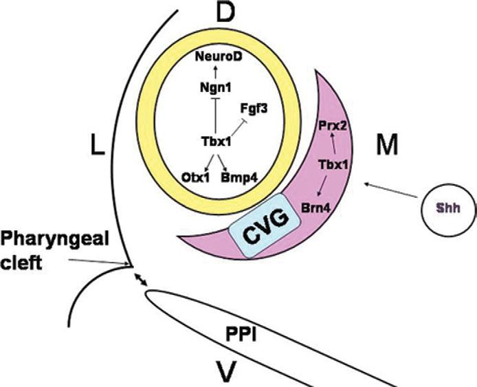
Most 22q11.2 deletion syndrome (22q11DS) patients have middle and outer ear anomalies, whereas some have inner ear malformations. Tbx1, a gene hemizygously deleted in 22q11DS patients and required for ear development, is expressed in multiple tissues during embryogenesis. To determine the role of Tbx1 in the first pharyngeal pouch (PPI) in forming outer and middle ears, we tissue-specifically inactivated the gene using Foxg1-Cre. In the conditional mutants, PPI failed to outgrow, preventing the middle ear bone condensations from forming. Tbx1 was also inactivated in the otic vesicle (OV), resulting in the failure of inner ear sensory organ formation, and in duplication of the cochleovestibular ganglion (CVG). Consistent with the anatomical defects, the sensory genes, Otx1 and Bmp4 were downregulated, whereas the CVG genes, Fgf3 and NeuroD, were upregulated. To delineate Tbx1 cell-autonomous roles, a more selective ablation, exclusively in the OV, was performed using Pax2-Cre. In contrast to the Foxg1-Cre mutants, Pax2-Cre conditional mutant mice survived to adulthood and had normal outer and middle ears but had the same inner ear defects as the Tbx1 null mice, with the same gene expression changes. These results demonstrate that Tbx1 has non-cell autonomous roles in PPI in the formation of outer and middle ears and cell-autonomous roles in the OV. Periotic mesenchymal markers, Prx2 and Brn4 were normal in both conditional mutants, whereas they were diminished in Tbx1-/- embryos. Thus, Tbx1 in the surrounding mesenchyme in both sets of conditional mutants cannot suppress the defects in the OV that occur in the null mutants.


Figures

Figure 1
Figure 1
(A) Generation of the Tbx1 conditional mutant mice. Genomic structure of the Tbx1 WT locus from exon 1 to 4 is shown on top. Standard gene targeting techniques were used to generate the Tbx1 hygro allele by inserting a single loxP site between exons 1 and 2 and a Hygromycin resistance cassette between exons 3 and 4. The Tbx1hygro/ + mice were mated with Ldh1-Cre mice to delete the Hygromycin resistance cassette and generate the Tbx1 flox/+ mice. Tbx1 floxed mice were crossed to Foxg1-Cre and Pax2-Cre mice to produce the Tbx1 conditional mutant mice, containing the Tbx1 null allele shown on the right. (B) Strategy to generate tissue-specific Tbx1 mutants. In WT embryos Tbx1 is expressed in both the OV and the surrounding PM, as well as in the PPI. Tbx1 expression is shown in blue. The Foxg1-Cre (left) and Pax2-Cre (right) strains mediate Tbx1 inactivation in the OV (shaded grey) while the PM expression domain remains intact. The Foxg1-Cre strain also mediates inactivation of Tbx1 in the PPI (shown in shaded grey). PCI, first pharyngeal cleft; Foxg1-Cre KO, Tbx1 null/flox;Foxg1-Cre/+, Pax2-Cre KO, Tbx1 null/flox; Pax2-Cre tg.
Figure 2
Figure 2
Tbx1 expression in E9.5 control embryos shown by IHC on sagittal sections with a Tbx1 polyclonal antibody (A) and by whole mount ISH with Tbx1 (B). Note Tbx1 expression in the OV, the pharyngeal pouches (PP, asterisks) and in the periotic mesenchyme (PM) and the core mesoderm of the pharyngeal arches (CM) in (A and B). Foxg1-Cre (C) and Pax2-Cre (E) activity shown by x-gal staining (lacZ) on E9.5 sagittal sections of Foxg1-Cre/ROSA26 (R26) and Pax2-Cre/ROSA26 (R26) progeny. Both Cre strains are active in the OV, while the Foxg1-Cre strain is also active in the pharyngeal pouches (stars in C). (D and F) Tbx1 expression by ISH in E9.5 Tbx1 conditional null mutant embryos, generated with the Foxg1-Cre strain (Foxg1-Cre KO) (D) and with the Pax2-Cre strain (Pax2-Cre KO) (F). Tbx1 is inactivated in the OV of both sets of mutants (OV in D and F), while the core mesoderm (CM) expression domain is still present. Tbx1 expression by IHC with a polyclonal Tbx1 antibody on E9.5 control (G) and Pax2-Cre KO (H). Note Tbx1 expression in the pharyngeal pouches (asterisks), and in the core mesoderm (CM). G and H are sagittal sections. ISH with Tbx1 on E9.5 sagittal sections of control (I) and Foxg1-Cre KO embryos (J). Note that Tbx1 expression is gone from the OV, and pharyngeal pouches (asterisks) of the Foxg1-Cre KO embryo (J). ISH with Tbx1 on E9.0 control (K) and Pax2-Cre KO mutant (L). Tbx1 expression is evident in the ventral OV in the control (K), but is absent in the Pax2-Cre KO mutant (L). The OV in the Pax2-Cre KO mutant (L) has been outlined with a red dashed line. Tbx1 is still present in the pharyngeal pouches (asterisks) of the mutant (L). Foxg1-Cre KO, Tbx1 null/flox; Foxg1-Cre/+, Pax2-Cre KO, Tbx1 null/flox; Pax2-Cre tg.
Figure 3
Figure 3
E10.5 transverse histological sections of control (A), Foxg1-KO (B) and Pax2-Cre KO (C) embryos. The invaginating pharyngeal endoderm in control (A) and Pax2-Cre KO (C) embryos is denoted with arrowheads. E17.5 transverse histological sections of control (D and E), Foxg1-Cre KO conditional mutants (F and G) and Pax2-Cre KO (H and I) embryos. Note the presence of the nascent tympanum in controls (E) and Pax2-Cre KOs (I), denoted by an arrow, and the middle ear bones (M), as opposed to Foxg1-Cre KOs (F,G). Horizontal histological sections of adult conditional mutant Pax2-Cre KO (J) and control (K) mice show the presence of normal middle ear structures in mutants (J). Sagittal histological sections of E10.5 control (L), Foxg1-Cre (M) and Pax2-Cre (N) mutant embryos. Early OV development is normal in both sets of mutant embryos, but the structure is slightly hypoplastic by E10.5 (M and N). The CVG is also enlarged in both Foxg1-Cre (M) and Pax2-Cre (N) mutants, when compared with a control embryo (L). CO, cochlea, SC, semicircular canals, P, pinna, O, ossicles (middle ear), Control, Tbx1+/−, Foxg1-Cre KO, Tbx1 null/flox; Foxg1-Cre/+, Pax2-Cre KO, Tbx1 null/flox; Pax2-Cre tg.
Figure 4
Figure 4
Development of the CVG in E10.5–E11.5 control (A and B), Foxg1-Cre KO (C and D) and Pax2-Cre KO (E and F) embryos. Whole-mount ISH with a NeuroD probe on E10.5 embryos (A, C and E). IHC with a monoclonal m2H3 anti-neurofilament antibody on E11.5 sagittal sections (B, D and F). Control, Tbx1+/−, Foxg1-Cre KO, Tbx1 null/flox; Foxg1-Cre, Pax2-Cre KO, Tbx1 null/flox; Pax2-Cre tg.
Figure 5
Figure 5
Molecular marker analysis by ISH. Fgf3 expression in control (A), Foxg1-Cre (B) and Pax2-Cre (C) mutant embryos at E10.5. Expression of Otx1 in E10.5 control (D), Foxg1-Cre (E) and Pax2-Cre (F) mutant embryos. Bmp4 expression in control (G), Foxg1-Cre (H) and Pax2-Cre mutant embryos (I). Control, Tbx1+/−, Foxg1-Cre KO, Tbx1 null/flox; Foxg1-Cre/+, Pax2-Cre KO, Tbx1 null/flox; Pax2-Cre tg.
Figure 6
Figure 6
PM specification shown by expression of marker genes. Prx2 expression is shown by ISH on E10.5 control (A), Foxg1-Cre KO (B) and Pax2-Cre KO (C) embryos. The arrow points to the PM expression domain. Brn4 expression by IHC with a polyclonal Brn4 antibody on control (D and E), Tbx1 null/flox; Foxg1-Cre/+ (F and G), Tbx1 null/flox; Pax2-Cre tg (H and I) and Tbx1−/− (J and K) sagittal sections of E11.5 embryos. Control, Tbx1+/−, Foxg1-Cre KO, Tbx1 null/flox; Foxg1-Cre/+, Pax2-Cre KO, Tbx1 null/flox; Pax2-Cre tg.
Figure 7
Figure 7
Model of Tbx1 function in ear development. The inner ear develops from the ectodermally derived OV, shown in yellow. Adjacent to the OV is the PM (pink). Tbx1 in the OV plays a dual role in inner ear development by inducing sensory organ formation in the posterior OV through induction of Otx1 and Bmp4 and by suppressing neurogenesis in the anterior OV through negative regulation of Ngn1 and NeuroD. The suppression of neurogenesis leads to the presence of an expanded CVG rudiment, shown in blue. In parallel, Tbx1 in the PM plays a role in mesenchymal tissue specification through induction of Brn4 and Prx2. This process might be mediated by Shh signaling from the notochord (circle) and the floor plate. Tbx1 in the PPI mediates tissue interactions between the endoderm of the pouches and the ectoderm of the first pharyngeal cleft to promote middle and outer ear development (double-headed arrows).

References

    1. DiGeorge A. A new concept of the cellular basis of immunity. J. Pediatr. 1965;67:907.
    1. Emanuel BS, McDonald-McGinn D, Saitta SC, Zackai EH. The 22q11.2 deletion syndrome. Adv. Pediatr. 2001;48:39–73. - PubMed
    1. McDonald-McGinn DM, Kirschner R, Goldmuntz E, Sullivan K, Eicher P, Gerdes M, Moss E, Solot C, Wang P, Jacobs I, et al. The Philadelphia story: the 22q11.2 deletion: report on 250 patients. Genet. Couns. 1999;10:11–24. - PubMed
    1. Greenberg F. What defines DiGeorge anomaly? J. Pediatr. 1989;115:412–413. - PubMed
    1. Schuknecht HF. Mondini dysplasia: a clinical and pathological study. Ann. Otol. Rhinol. Laryngol. Suppl. 1980;89:1–23. - PubMed

Publication types