. 2006 Aug;20(8):1796-809.
doi: 10.1210/me.2005-0327. Epub 2006 Apr 6.

Thyroid-specific enhancer-binding protein/NKX2.1 is required for the maintenance of ordered architecture and function of the differentiated thyroid

Affiliations

Thyroid-specific enhancer-binding protein/NKX2.1 is required for the maintenance of ordered architecture and function of the differentiated thyroid

Takashi Kusakabe et al. Mol Endocrinol. 2006 Aug.

Abstract

Thyroid-specific enhancer-binding protein (T/ebp)/Nkx2.1-null mouse thyroids degenerate by embryonic day (E) 12-13 through apoptosis whereas T/ebp/Nkx2.1-heterogyzgous mice exhibit hypothyroidism with elevated TSH levels. To understand the role of T/ebp/Nkx2.1 in the adult thyroid, a thyroid follicular cell-specific conditional knockout (KO) mouse line, T/ebp(fl/fl);TPO-Cre, was established that expresses Cre recombinase under the human thyroid peroxidase (TPO) gene promoter. These mice appeared to be healthy and exhibited loss of T/ebp/Nkx2.1 expression in many, but not all, thyroid follicular cells as determined by immunohistochemistry and real-time PCR, thus presenting a T/ebp-thyroid-conditional hypomorphic mice. Detailed analysis of the thyroids from T/ebp(fl/fl), T/ebp(fl/fl);TPO-Cre, and T/ebp(fl/ko) mice, where the latter mouse line is derived from crosses with the original T/ebp/Nkx2.1-heterozygous mice, revealed that T/ebp(fl/fl);TPO-Cre mice can be classified into two groups with different phenotypes: one having atrophic/degenerative thyroid follicles with frequent presence of adenomas and extremely high serum TSH levels, and the other having an altered thyroid structure with reduced numbers of extraordinary dilated follicles consisting of excessive numbers of follicular cells as compared with those usually found in the normal thyroid. The latter phenotype was also observed in aged T/ebp(fl/ko) mouse thyroids. In vitro three-dimensional thyroid primary cultures using thyroids from T/ebp(fl/fl);TPO-Cre, T/ebp(fl/ko), and T/ebp(fl/fl) mice, and the latter treated with recombinant adenovirus with and without Cre expression, demonstrated that only cells from T/ebp(fl/fl) mice without adeno-Cre treatment formed follicular structures. Taken together, these results suggest that T/ebp/Nkx2.1 is required for maintenance of the normal architecture and function of differentiated thyroids.


Figures
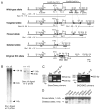
Fig. 1
Fig. 1. Generation of the T/ebp-Floxed Mouse
A, Diagram of the T/ebp gene (wild-type allele), targeted allele, floxed allele, deleted allele, and the original KO allele. The targeting vector was composed of EcoRI-EcoRI and EcoRI-EcoRV fragments, shown on the top. Shaded and open boxes represent coding and noncoding regions of the gene, respectively, and exons are numbered. The neomycin cassette is shown by an open box marked with pGK-Neo for the targeted allele and the black box marked with Neo for the original KO allele. Solid triangles represent the loxP sites. The probes (5′-probe and 3′-probe) used to assess recombination events and the expected lengths of restriction enzyme fragments are indicated. The locations of PCR primers (5neo/3neo and 5KO/3KO) used for genotyping are also shown. Restriction enzyme sites: B, BamHI; RI, EcoRI; RV, EcoRV; H, HindIII; Ns, NsiI; Sc, SacI; St, StuI; Xh, XhoI. B, Southern blot analysis of homologous recombination in ES cells electroporated with the targeting vector. Genomic DNAs were digested with StuI or BamHI and hybridized with a 3′-probe or 5′-probe, respectively. The StuI-digested DNA analyzed with the 3′-probe detected approximately 14 and 1 kb bands for wild-type allele (+), and 3 and 1 kb bands for targeted allele (t), whereas BamHI-digested DNA analyzed with the 5′-probe produced 11- and 8-kb bands for wild-type and targeted alleles, respectively. C, PCR analysis of mouse tail DNA. Left panel: Floxed allele (fl) was distinguished from wild-type or KO (ko) allele by primer set 5neo/3neo. Floxed allele was represented by an amplified 220-bp band, whereas wild-type or KO allele produced a 540-bp band. Right panel: The KO allele was further distinguished from wild-type or floxed allele by primer set 5KO/3KO, which amplified a 220-bp band only for the KO allele. D, Southern blot analysis for deletion of the floxed T/ebp gene in various organs from 1-month-old T/ebp(fl/fl); TPO-Cre mice. Genomic DNAs (5 μg) from eight different tissues were digested with StuI, subjected to electrophoresis, and hybridized with 32P-labeled 3′-probe. Ex, Exon.
Fig. 2
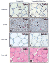
Fig. 2. Partial Loss of T/ebp Protein Expression in T/ebp(fl/fl); TPO-Cre Mouse Thyroids
Thyroids from 1-month-old or E16.5 embryos of T/ebp(fl/fl) or T/ebp(fl/fl); TPO-Cre mice were subjected to T/ebp immunostaining, TUNEL assay, or hematoxylin and eosin (H&E). T/ebp immunostaining and TUNEL assay of 1-month-old thyroids were performed on serially prepared sections. A and B, In control T/ebp(fl/fl) mice, T/ebp is highly expressed in most follicular cells of 1-month-old mice (panel A: representative is shown by arrow). In T/ebp(fl/fl); TPO-Cre mouse thyroids, the expression of T/ebp is frequently lost (panel B: representatives are shown by arrowheads). C and D, T/ebp is highly expressed (representative is shown by arrow), weakly expressed (representative is shown by arrowhead), or not expressed (representative is shown by red arrow) in both T/ebp(fl/fl) and T/ebp(fl/fl); TPO-Cre E16.5 thyroids. E and F, TUNEL analysis shows no evidence of apoptotic degeneration in either 1-month-old T/ebp(fl/fl) or T/ebp(fl/fl); TPO-Cre mouse thyroids. G and H, Hematoxylin and eosin staining of 1-month-old adult thyroid glands revealed no gross histological differences between T/ebp(fl/fl) and T/ebp(fl/fl); TPO-Cre mouse thyroids. Magnification, ×200. mo, Month.
Fig. 3
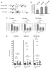
Fig. 3. Analyses of the Recombination Frequency of the T/ebp-Floxed Allele, T/ebp Expression Levels, and Serum TSH Levels
A, Schematic diagram of the location of primers P1, P2, and P3 (arrows) that were used for determination of recombination frequency, and primers a and b (shaded arrowheads) that were used for quantitation of T/ebp mRNA levels by real-time PCR. The thick line indicates the sequence in between two XhoI sites that is present only in the floxed allele derived from the targeting vector and differs from that of KO allele. The P2/P3 primer pair generates an amplicon (164 bp) only from the unrecombined floxed allele, but not from the recombined deleted allele because of the loss of the P2 primer binding sequence following Cre/loxP excision of exon2, nor the KO allele because P2 primer sequence does not exist. The P1/P3 primer pair generates an amplicon (165 bp) from the recombined deleted allele, but not from the unrecombined floxed or KO allele because of the large amplicon size. The diagram is not drawn to scale. Restriction enzyme sites: Xh, XhoI. B, Frequency of T/ebp-floxed gene recombination in T/ebp(fl/fl); TPO-Cre mice. The ratio of the recombined allele is shown as a percentage of the total floxed (unrecombined and recombined together) alleles, which was set at 100%. Values are the means ± SE. C, Expression levels of T/ebp mRNA in thyroid glands. All mRNA levels are normalized to 18S rRNA and presented as a relative ratio to the level in T/ebp(fl/fl) mice in each age group set to 1.0. Values are the means ± SE. D, Serum TSH levels in T/ebp(fl/fl), T/ebp(fl/fl); TPO-Cre, and T/ebp(fl/ko) mice. Each dot represents a TSH level for single mouse, and horizontal bars represent the mean values. Asterisks indicate mice that demonstrated histological atrophic/degenerative abnormalities in their thyroids as described in the text. NS, Statistically not significant; mo, month.
Fig. 4
Fig. 4. Histological Abnormalities of T/ebp(fl/fl); TPO-Cre Conditional Hypomorphic Mouse Thyroids
Hematoxylin and eosin staining of transverse sections of thyroids from 10-month-old T/ebp(fl/fl) control (A) and T/ebp(fl/fl); TPO-Cre conditional hypomorphic mice (B and C). Tr, Trachea. B, Severely impaired thyroid gland commonly seen among mice that have highly elevated serum TSH levels. Size of the gland is significantly smaller as compared with that of control thyroid gland shown in panel A. C, Cystic phenotype of the thyroid gland, which is frequently observed in T/ebp(fl/fl); TPO-Cre and T/ebp(fl/ko) mouse lines, particularly as they age. Dilated follicles preferentially reside near the periphery of the lobe. D, Higher magnification of severely affected thyroid gland common to mice that have highly elevated serum TSH levels. The lobe consists of intact follicles (representatives are shown by red asterisks), degenerative follicles with complete or partial colloid depletion (representatives are shown by black asterisks), and multiple small follicles (arrows) that intervene between intact and degenerative follicles. E, High-power view of degenerative follicle. Desquamative thyrocyte (arrowhead) and cell debris (arrow) in depleted colloid are seen in the follicular lumen. Follicular cell lining is partly disappeared (representatives are shown by asterisks). F, Follicular adenoma (lower left part of the panel), which is frequently seen in mice with high TSH levels, is well demarcated from adjacent degenerative follicles (upper right). G–I, Immunostaining for T/ebp. Follicular cells in adenoma highly express T/ebp (G, representatives are shown by black asterisks), whereas the follicles in which lining thyrocytes have lost T/ebp expression become degenerative (G, representatives are shown by red asterisks), and sometimes contain cell debris and desquamative cells in the lumen (H, arrows). I, Almost all follicular cells in dilated follicles as seen in panel C including those from T/ebp(fl/fl); TPO-Cre mice clearly express T/ebp. A part of the follicles is enlarged (inset). Magnification: A–C, ×10; D, ×100; F, G, and I, ×200; E and H, ×400.
Fig. 5
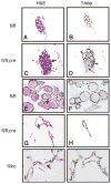
Fig. 5. Three-Dimensional Thyroid Primary Cultures in Collagen Gels
Seven-day culture of thyroid follicles (A–D), or 3-wk culture of individual thyroid follicular cells (E–J) obtained from T/ebp(fl/fl), T/ebp(fl/fl); TPO-Cre, and T/ebp(fl/ko) mouse thyroids. Paraffin sections were subjected to histological analysis using hematoxylin and eosin or T/ebp immunohistochemistry. A and B and C and D are serially prepared sections. T/ebp(fl/fl) mouse follicles maintained round shape with colloid in the lumen (A) that expressed T/ebp (B). T/ebp(fl/fl); TPO-Cre follicular cultures consisted of a mixture of follicles that were different in size and shape from round (C, representative is shown by red arrowhead) to oval to irregular (C, representatives are shown by black arrowheads), and the levels of colloid in the lumen from normal to none (C, the latter representative is shown by arrow). Some follicles exhibited T/ebp expression (D, arrowhead) whereas others had no T/ebp expression, particularly in the area where most follicles were obliterated, exhibiting solid growth pattern (C and D, shown by thin arrow). Single follicular cell grew to a cluster of spherical follicles in T/ebp(fl/fl) with a plenty of colloid in the lumen (E, representative is shown by arrow) whereas T/ebp(fl/fl); TPO-Cre and T/ebp(fl/ko) follicular cell produced irregular shaped polygonal follicles with no colloid accumulation (G and I, representative is shown by arrow). Most follicular cells from T/ebp(fl/fl) and T/ebp(fl/ko) mice were positive for T/ebp expression (F and J) whereas those of T/ebp(fl/fl); TPO-Cre mice exhibited various degrees of T/ebp expression (H). Experiments were separately carried out at least three times for each culture condition and genotype, and each time the same results were obtained. Magnification: A–D, ×100, E–J, ×200.
Fig. 6
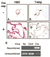
Fig. 6. Effect of the Loss of T/ebp on Structure of T/ebp(fl/fl) Thyroid in Three-Dimensional Thyroid Primary Cultures
Individual thyrocytes isolated from T/ebp(fl/fl) thyroids were treated with recombinant adenovirus containing vector only as control (A and B) or those expressing Cre recombinase (C and D), followed by three-dimensional culture in collagen. Two weeks later, the cultures were subjected to histological analysis with hematoxylin and eosin (A and C) or T/ebp immunohistochemistry (B and D). Round follicles started appearing in control cultures with a weak pink color, indicative of colloid in the lumen (A) and strong T/ebp expression in most of cells (B, representatives are shown by arrow) whereas no follicular structures were obtained from Cre-expressing adenovirus-treated thyroid cells (C), most of which had weak (D, representatives are shown by arrowhead) or no expression of T/ebp (D, representatives are shown by red arrowhead), and only a few cells showed strong positive staining (D, shown by arrow). Experiments were carried out using thyroids from three independent T/ebp(fl/fl) mice, and the same results were obtained for all three. Magnification, ×200. E, Genomic PCR analysis of recombined (164 bp) and unrecombined T/ebp allele (165 bp) using DNAs isolated from control cells without any virus treatment (No), cells treated with control virus (Cont), and cells treated with adenovirus expressing Cre recombinase (Cre). Only the latter DNA produced a recombined band. exp, Experiment; H&E, hematoxylin and eosin.

Similar articles

Cited by

References

    1. Kaufman M, Bard J. The anatomical basis of mouse development. London: Academic Press; 1999.
    1. Manley NR, Capecchi MR. Hox group 3 paralogs regulate the development and migration of the thymus, thyroid, and parathyroid glands. Dev Biol. 1998;195:1–15. - PubMed
    1. Di Lauro R, De Felice M. Thyroid gland: anatomy and development. In: DeGroot L, Jameson J, editors. Endocrinology. Philadelphia: Saunders; 2001. pp. 1268–1278.
    1. Missero C, Cobellis G, De Felice M, Di Lauro R. Molecular events involved in differentiation of thyroid follicular cells. Mol Cell Endocrinol. 1998;140:37–43. - PubMed
    1. Kusakabe T, Hoshi N, Kimura S. Origin of the ultimobranchial body cyst: T/ebp/Nkx2.1 expression is required for development and fusion of the ultimobranchial body to the thyroid. Dev Dyn. 2006;235:1300–1309. - PMC - PubMed

Publication types

MeSH terms