. 2006 Apr 18;103(16):6172-7.
doi: 10.1073/pnas.0601686103. Epub 2006 Apr 6.

Chaperone-mediated assembly of centromeric chromatin in vitro

Affiliations

Chaperone-mediated assembly of centromeric chromatin in vitro

Takehito Furuyama et al. Proc Natl Acad Sci U S A. .

Abstract

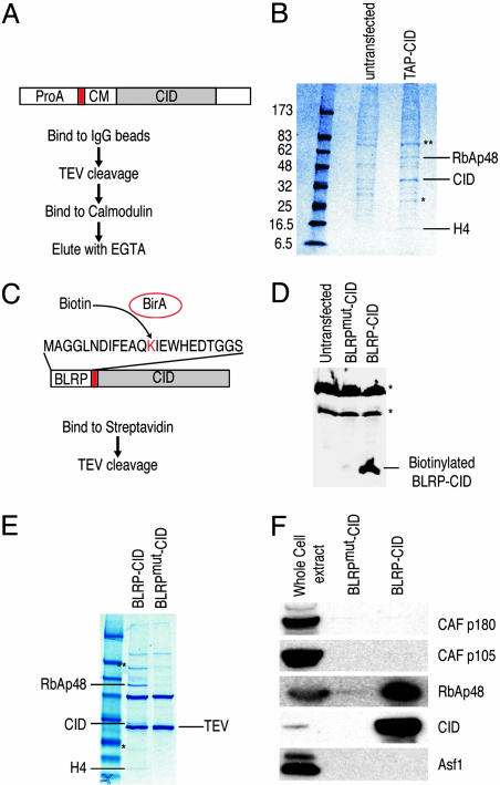
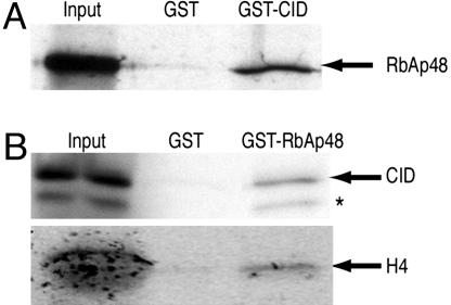
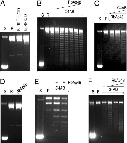
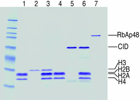
Every eukaryotic chromosome requires a centromere for attachment to spindle microtubules for chromosome segregation. Although centromeric DNA sequences vary greatly among species, centromeres are universally marked by the presence of a centromeric histone variant, centromeric histone 3 (CenH3), which replaces canonical histone H3 in centromeric nucleosomes. Conventional chromatin is maintained in part by histone chaperone complexes, which deposit the S phase-limited (H3) and constitutive (H3.3) forms of histone 3. However, the mechanism that deposits CenH3 specifically at centromeres and faithfully maintains its chromosome location through mitosis and meiosis is unknown. To address this problem, we have biochemically purified a soluble assembly complex that targets tagged CenH3 to centromeres in Drosophila cells. Two different affinity procedures led to purification of the same complex, which consists of CenH3, histone H4, and a single protein chaperone, RbAp48, a highly abundant component of various chromatin assembly, remodeling, and modification complexes. The corresponding CenH3 assembly complex reconstituted in vitro is sufficient for chromatin assembly activity, without requiring additional components. The simple CenH3 assembly complex is in contrast to the multisubunit complexes previously described for H3 and H3.3, suggesting that centromeres are maintained by a passive mechanism that involves exclusion of the complexes that deposit canonical H3s during replication and transcription.


Conflict of interest statement

Conflict of interest statement: No conflicts declared.

Figures

Fig. 1.
Fig. 1.
Purification of the CID assembly complex from S2 cells. (A) Purification scheme using TAP-tagged CID. ProA, protein A domain; red box, TEV cleavage site; CM, calmodulin-binding peptide. (B) TAP-CID-associated complex was purified and separated on a gradient gel. Mock purification was performed with extracts from untransfected cells. After Coomassie blue staining, protein bands that are only present in the TAP-CID lane were excised and identified by MS. (C) Biotin-mediated purification scheme. Coexpressed E. coli biotin ligase (BirA) transfers biotin to the lysine residue of BLRP tag. Red box, TEV cleavage site. A point mutation in BLRP (BLRPmut-CID) that changes K to R, rendering it nonbiotinylatable, was also made (not shown). (D) Western blot analysis with streptavidin-horseradish peroxidase on untransfected, BLRPmut-CID, and BLRP-CID cells. Asterisks indicate endogenously biotinylated proteins in the cell. Western blots with anti-CID antibody detect both BLRP-CID and BLRPmut-CID equally (data not shown). (E) The BLRP-CID-associated complex contains the same proteins found in the TAP-purified complex. In B and E, ∗ indicates partially degraded CID, and ∗∗ indicates a protein enriched but present in the mock-purified material. (F) Western blot analysis was performed on samples shown in the Coomassie blue-stained gel (E), with various antibodies confirming the identities of RbAp48 and CID. There are no detectable levels of other subunits of CAF-1 complex (p180 and p105) or Asf1 in the purified material.
Fig. 2.
Fig. 2.
BLRP-CID localizes specifically to centromeres. (A and B) Cytological detection of BLRP-CID by streptavidin-Alexa Fluor 488 (green) on interphase (A) or mitotic (B) chromosomes of S2 cells (DAPI shown in blue) shows a characteristic centromere-staining pattern with only a faint noncentromeric signal. (C) No specific staining of BLRPmut-CID cells is seen. Immunological methods are described in Supporting Materials and Methods.
Fig. 3.
Fig. 3.
RbAp48 and CID directly interact in vitro. (A) A GST-CID fusion protein binds 35S-labeled RbAp48 translated in vitro. (B) A GST-RbAp48 fusion protein binds 35S-labeled CID and H4 translated in vitro. Some CID is either degraded or prematurely terminated (∗). An abundant protein present in the in vitro translation reaction (Promega) comigrates with the in vitro translated H4 in the input lane, causing a broad diffuse appearance. Input corresponds to 20% of the material used for binding assays. GST pull-down is described in Supporting Materials and Methods.
Fig. 4.
Fig. 4.
Analysis of nucleosome assembly by plasmid supercoiling assays. Supercoiled pCR4–360 × 8 plasmid was purified from E. coli (S) and relaxed by addition of topoisomerase I (R). (A) Chromatin assembly was performed by incubating the relaxed plasmid with the indicated protein complexes purified from S2 cells. (B) CID chromatin assembly reactions were performed by incubating the relaxed plasmid with CID/H4/H2A/H2B (recombinant CID and HPLC-purified H4/H2A/H2B) and increasing amounts of recombinant RbAp48. −, no RbAp48 added. (C) Same as in B except that the amount of RbAp48 was fixed, and increasing amounts of C4AB were added to the reaction. (D) A control assembly reaction with only RbAp48 does not yield any supercoils. (E) A plasmid supercoiling assay on pUC19 is markedly less efficient compared with pCR4–360 × 8. (F) Same as in B except that H3 was substituted for CID.
Fig. 5.
Fig. 5.
Reconstitution of the CID assembly complex from purified components. In vitro reconstituted CID assembly complex components: lanes 1 and 4, HPLC-purified histone H4/H2A/H2B; lane 2, HPLC-purified H3; lane 3, refolded H3/H4/H2A/H2B; lane 5, recombinant CID; lane 6, refolded CID/H4/H2A/H2B; lane 7, recombinant RbAp48.
Fig. 6.
Fig. 6.
Analysis of in vitro assembled CID nucleosomes. (AD) EM images of naked DNA before assembly into chromatin (A), circular DNA assembled into CID chromatin (B), and linear DNA assembled into CID chromatin and diluted into physiological salt (C) or assembly buffer (D) before imaging. Sample grids were rotary-shadowed. Images are representative of ≈100 molecules counted during three independent assembly reactions. EM methods are described in Supporting Materials and Methods. (E) DNase I digestion of in vitro assembled CID chromatin. A ladder of ≈10-bp periodicity indicates that DNA had been wrapped around histones. M, 10-bp ladder marker.

References

    1. Choo K. H. The Centromere. London: Oxford Univ. Press; 1997.
    1. Malik H. S., Henikoff S. Curr. Opin. Genet. Dev. 2002;12:711–718. - PubMed
    1. Amor D. J., Choo K. H. Am. J. Hum. Genet. 2002;71:695–714. - PMC - PubMed
    1. Nagaki K., Cheng Z., Ouyang S., Talbert P. B., Kim M., Jones K. M., Henikoff S., Buell C. R., Jiang J. Nat. Genet. 2004;36:138–145. - PubMed
    1. Henikoff S., Ahmad K., Malik H. S. Science. 2001;293:1098–1102. - PubMed

Publication types

MeSH terms

LinkOut - more resources