. 2006 Apr 18;103(16):6404-9.
doi: 10.1073/pnas.0510283103. Epub 2006 Apr 6.

Phosphorylation and activation of PINOID by the phospholipid signaling kinase 3-phosphoinositide-dependent protein kinase 1 (PDK1) in Arabidopsis

Affiliations

Phosphorylation and activation of PINOID by the phospholipid signaling kinase 3-phosphoinositide-dependent protein kinase 1 (PDK1) in Arabidopsis

Hicham Zegzouti et al. Proc Natl Acad Sci U S A. .

Abstract

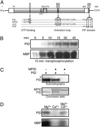
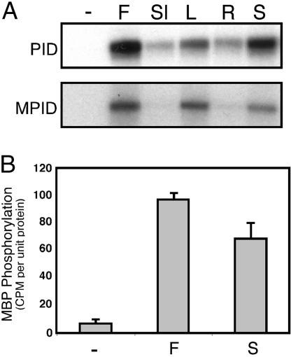
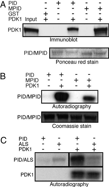
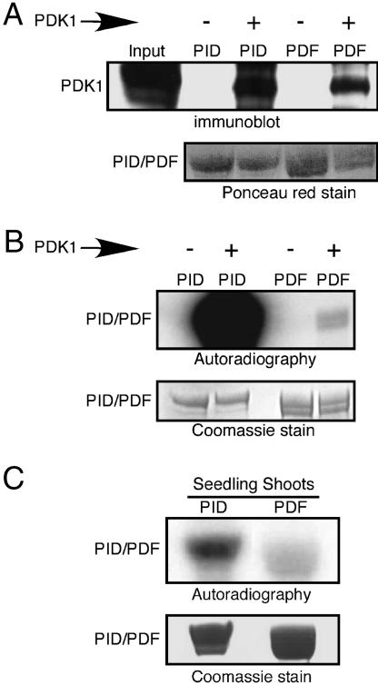
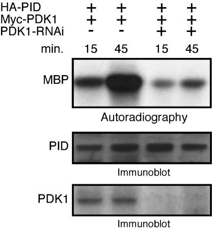
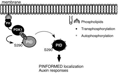
Activity of the serine-threonine protein kinase PINOID (PID) has been implicated in the asymmetrical localization of the membrane-associated PINFORMED (PIN) family of auxin transport facilitators. However, the means by which PID regulates PIN protein distribution is unknown. We have used recombinant PID protein to dissect the regulation of PID activity in vitro. We demonstrate that intramolecular PID autophosphorylation is required for the ability of PID to phosphorylate an exogenous substrate. PID-like mammalian AGC kinases act in a phosphorylation cascade initiated by the phospholipid-associated kinase, 3-phosphoinositide-dependent protein kinase 1 (PDK1), which binds to the C-terminal hydrophobic PDK1-interacting fragment (PIF) domain found in PDK1 substrates. We find that Arabidopsis PDK1 interacts with PID, and that transphosphorylation by PDK1 increases PID autophosphorylation. We show that a PID activation loop serine is required for PDK1-dependent PID phosphorylation. This activation is rapid and requires the PIF domain. Cell extracts from flowers and seedling shoots dramatically increase PID phosphorylation in a tissue-specific manner. A PID protein variant in which the PIF domain was mutated failed to be activated by the seedling shoot extracts. PID immunoprecipitated from Arabidopsis cells in which PDK1 expression was inhibited by RNAi showed a dramatic reduction in transphosphorylation of myelin basic protein substrate. These results indicate that AtPDK1 is a potent enhancer of PID activity and provide evidence that phospholipid signaling may play a role in the signaling processes controlling polar auxin transport.


Conflict of interest statement

Conflict of interest statement: No conflicts declared.

Figures

Fig. 1.
Fig. 1.
Intramolecular autophosphorylation activates PID transphosphorylation activity. (A) Schematic representation of PINOID, showing the wild-type sequence of the ATP binding domain, activation loop, and PIF domain, and the corresponding changes introduced in the MPID, ALS, and PDF derivatives. (B) Autoradiograph showing that autophosphorylation of PID is required for its transphosphorylation of MBP. (Upper) PID was incubated in kinase buffer for the indicated times. (Lower) For each time point, unlabeled, autophosphorylated PID was mixed with MBP and 5 μCi of [γ-32P]ATP (1Ci = 37 GBq) and incubated for an additional 15 min. The results shown are representative of three independent experiments. (C) PID autophosphorylates intramolecularly. His-tagged PID and GST-tagged MPID were coincubated with 10 μCi of [γ-32P]ATP and kinase buffer. The asterisk marks the location of nonphosphorylated GST-MPID visible in the Coomassie-stained gel. All autoradiographs shown are 24-h exposures. Identical results were obtained in two independent experiments. (D) Calcium inhibition of PID activity. PID activity was assayed in the presence of 15 mM MgCl2, 15 mM CaCl2, or both. For each reaction, PID was allowed to autophosphorylate (Upper) for 45 min before the addition of 3 μg of MBP (Lower). Experiments were performed in triplicate.
Fig. 2.
Fig. 2.
Transactivation of PID by tissue-specific cell extracts. (A) Total protein purified from mixed stage flowers (F), siliques (Sl), rosette leaves (L), 10-day-old seedling shoots (S), or roots (R) was incubated with GST:PID or GST:MPID. PID labeling was detected by autoradiography after PAGE. (B) GST-PID was phosphorylated by tissue-specific protein extracts as described in A, washed, and incubated with MBP to assess PID activity. Incorporation of γ-32P by MBP was quantified by using a liquid scintillation counter. Data are representative of three independent experiments; error bars represent standard error. Autoradiographs were exposed for 12 h.
Fig. 3.
Fig. 3.
PID autophosphorylation is induced by PDK1 phosphorylation. (A) His:PDK1-containing bacterial lysate was incubated with GST, GST:PID, or GST:MPID bound to glutathione-agarose beads. Half of the sample was used to confirm PDK1 binding by Western blot analysis by using anti-polyhistidine antibody. (B) The remaining sample was used in a kinase assay in the presence of [γ-32P]ATP and subjected to PAGE. Incorporation of radiolabeled phosphate was determined by autoradiography. Binding and phosphorylation analyses were confirmed by three independent experiments. (C) An activation loop serine is required for PDK1 activation of PID. Wild-type and ALS mutant protein (see Fig. 1A) phosphorylation was analyzed in the absence or presence of PDK1 protein. Results shown were representative of two independent experiments. Autoradiographs were exposed for 1 h in B and C.
Fig. 4.
Fig. 4.
The PIF domain is required for PDK1-mediated PID phosphorylation in vitro and in vivo. (A) Binding of PDK1 to PID and PDF protein was visualized by Western blotting as described in the legend for Fig. 3. (B) PID phosphorylation level was compared with that of PDF in the absence or presence of bound PDK1. (C) Equivalent amounts of GST:PID or GST:PDF proteins were incubated with seedling shoot extract in kinase buffer in the presence of [γ-32P]ATP for 30 min. Protein phosphorylation was visualized by autoradiography. All experiments were performed in triplicate. Autoradiographs were exposed for 1 h in B and 12 h in C.
Fig. 5.
Fig. 5.
In vivo activation of PID by PDK1. PID was immunoprecipitated from cells cotransfected with HA-PID and Myc-PDK1 in the presence or absence of PDK1-RNAi. After PID autophosphorylation for the indicated times, MBP was added to the kinase reaction for an additional 30 min to asses PID transphosphorylation activity (Top). Equal amounts of proteins from the above transfected cells were used to confirm PID and PDK1 expression by immunoblotting by using HA (Middle) and Myc (Bottom) antibodies, respectively. Data are representative of two independent experiments.
Fig. 6.
Fig. 6.
Proposed role of PID in phospholipid-mediated membrane targeting. PDK1 is localized to specific membrane regions by binding through the pleckstrin homology (PH) domain to locally produced phosphoinositide ligands. PID is recruited to the membrane through the interaction of PDK1 with the PID PIF domain. PDK1 phosphorylation of Ser 290 within the activation loop stimulates PID autophosphorylation. Active PID phosphorylates substrate proteins, resulting in the targeted localization of the polar auxin transport complex machinery.

Similar articles

Cited by

References

    1. Friml J., Palme K. Plant Mol. Biol. 2002;49:273–284. - PubMed
    1. Gälweiler L., Guan C., Müller A., Wisman E., Mendgen K., Yephremov A., Palme K. Science. 1998;282:2226–2230. - PubMed
    1. Steinmann T., Geldner N., Grebe M., Mangold S., Jackson C. L., Paris S., Gälweiler L., Palme K., Jürgens G. Science. 1999;286:316–318. - PubMed
    1. Geldner N., Friml J., Stierhof Y. D., Jurgens G., Palme K. Nature. 2001;413:425–428. - PubMed
    1. Geldner N., Anders N., Wolters H., Keicher J., Kornberger W., Muller P., Delbarre A., Ueda T., Nakano A., Jürgens G. Cell. 2003;112:219–230. - PubMed

Publication types

MeSH terms