. 2006 Jun 9;281(23):16108-16.
doi: 10.1074/jbc.M600760200. Epub 2006 Apr 6.

Identification of a pH sensor in the furin propeptide that regulates enzyme activation

Affiliations

Identification of a pH sensor in the furin propeptide that regulates enzyme activation

Sylvain F Feliciangeli et al. J Biol Chem. .

Abstract

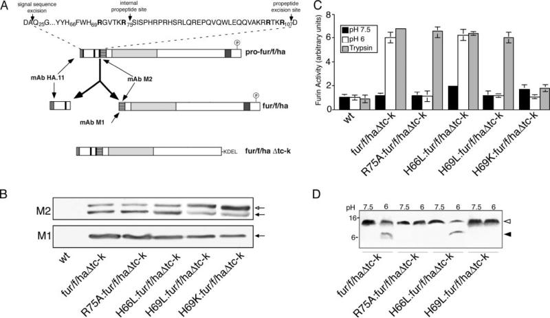
The folding and activation of furin occur through two pH- and compartment-specific autoproteolytic steps. In the endoplasmic reticulum (ER), profurin folds under the guidance of its prodomain and undergoes an autoproteolytic excision at the consensus furin site Arg-Thr-Lys-Arg107/ generating an enzymatically masked furin-propeptide complex competent for transport to late secretory compartments. In the mildly acidic environment of the trans-Golgi network/endosomal system, the bound propeptide is cleaved at the internal site 69HRGVTKR75/, unmasking active furin capable of cleaving substrates in trans. Here, by using cellular, biochemical, and modeling studies, we demonstrate that the conserved His69 is a pH sensor that regulates the compartment-specific cleavages of the propeptide. In the ER, unprotonated His69 stabilizes a solvent-accessible hydrophobic pocket necessary for autoproteolytic excision at Arg107. Profurin molecules unable to form the hydrophobic pocket, and hence, the furin-propeptide complex, are restricted to the ER by a PACS-2- and COPI-dependent mechanism. Once exposed to the acidic pH of the late secretory pathway, protonated His69 disrupts the hydrophobic pocket, resulting in exposure and cleavage of the internal cleavage site at Arg75 to unmask the enzyme. Together, our data explain the pH-regulated activation of furin and how this His-dependent regulatory mechanism is a model for other proteins.


Figures

FIGURE 1
FIGURE 1. Homology modeling of the furin propeptide
A, multiple sequence alignment between propeptides of the PC family. Predicted secondary structure of the furin propeptide and sequence conservation (black highlight, 100% similarity; dark gray highlight, >80% similarity, light gray highlight, >50% similarity) between PCs is depicted. Propep-tide residues are numbered in reference to the furin propeptide, which begins at Gln25 (18). B, ribbon representation of the furin propeptide structure obtained by homology modeling. His66 and His69 (blue) and Arg75 (red) are highlighted. C, surface representation of the propeptide-furin complex. The modeled propeptide (yellow) is docked onto the active site of furin (green). The internal propeptide cleavage site (red) and His69 (blue) are highlighted. D, surface representation of the secondary cleavage site illustrates His69 (blue) buried in the solvent-accessible pocket formed by Gly53, Phe54, Leu55, Phe67, and Trp68 hydrophobic residues (yellow).
FIGURE 2
FIGURE 2. His69 controls the pH-sensitive furin propeptide cleavage and enzyme activation in vitro
A, schematic representation of the furin constructs used in this study. Shown is full-length profurin containing the N-terminal prodomain with the excision and internal cleavage sites (black bars), the catalytic domain (light gray segment), the transmembrane domain (dark gray segment), and the cytosolic domain, which is phosphorylated by CK2 (circled P) to promote binding to PACS proteins (15, 17, 25). A FLAG epitope tag (horizontal bars) was inserted at the N terminus of mature furin such that the FLAG N terminus is exposed upon propeptide excision. mAb M2 recognizes either the blocked (profurin) or N-terminally exposed (mature furin) FLAG tag, whereas mAb M1 recognizes specifically the N-terminally exposed FLAG tag (mature furin). An HA epitope tag (vertical bars), which is recognized by mAb HA.11, was inserted at the N terminus of the prodomain. The sequence of the prodomain is shown at the top with the signal peptidase cleavage site as well as the propeptide excision and internal cleavage sites highlighted by vertical arrows. The P1/P4 Arg residues of the excision site and the P1/P6 Arg residues of the internal cleavage site are in boldface font. ER-localized, luminally restricted furin constructs (designated Δtc-k) were generated by replacing the transmembrane domain and cytosolic domains with the Lys-Asp-Glu-Leu (KDEL) ER localization signal. B, cell extracts from BSC-40 cells expressing a control viral vector (wt) or the indicated -Δtc-k ER-localized furin constructs were separated by SDS-PAGE and analyzed by Western blot using mAbs M1 or M2. Open arrow, profurin. Filled arrow, mature furin. C, membrane preparations from cells used in B were tested for furin activity using the Abz-Arg-Val-Lys-Arg-Gly-Leu-Ala-Tyr(NO2)-Asp-OH substrate, after incubation at pH 6.0 or 7.5 for 3 h, or at pH 7.5 for 1 h in the presence of trypsin followed by soybean trypsin inhibitor to block residual trypsin activity. Error bars represent the mean and S.E. of three independent experiments. D, membranes preparations from C were analyzed by 15% Tris-Tricine SDS-PAGE followed by Western blot using mAb HA.11. Mr values of the excised propeptide and the cleaved N-terminal propeptide fragment are indicated. Open arrowhead, intact propeptide. Filled arrowhead, cleaved propeptide.
FIGURE 3
FIGURE 3. The protonation state of His69 controls propeptide excision and enzyme activation in vivo
A, cell extracts from BSC-40 cells expressing a control viral vector (wt) or full-length epitope-tagged furin molecules containing the indicated mutations were separated by SDS-PAGE and analyzed by Western blot with mAbs M1 and M2. Profurin (open arrow) and mature furin (filled arrow) are indicated. B, crude membrane preparations of cells used in A were tested for furin activity at pH 7.5 using the Abz-Arg-Val-Lys-Arg-Gly-Leu-Ala-Tyr(NO2)-Asp-OH substrate peptide. Error bars represent the mean and S.E. of three independent experiments.
FIGURE 4
FIGURE 4. The protonation state of His69 affects the subcellular localization of furin
A, immunofluorescence microscopy of BSC-40 cells expressing fur/f/ha, H69L:fur/f/ha, or H69K:fur/ f/ha. Cells were fixed and stained with anti-furin PA1-062 and anti-TGN46 and then visualized with species-specific fluorescently labeled secondary antibodies. B, BSC-40 cells expressing fur/f/ha or H69L:fur/f/ha were pulse-labeled for 30 min with 100 μCi each of [3H]Arg and [3H]Leu and then chased with excess unlabeled Arg and Leu at 37 °C for the indicated times. Cell extracts were prepared, and mature furin molecules were immunoprecipitated with mAb M1, separated by 15% Tris-Tricine SDS-PAGE, and co-precipitating propeptide molecules (open arrowheads) were detected by fluorography and quantified by densitometry. C, immunofluorescence microscopy of BSC-40 cells expressing fur/f/ha or H69L:fur/f/ha. Cells were incubated with mAb M1 in the culture medium (30 μg/ml) for 1 h, and internalized mAb M1 was detected with a fluorescently labeled secondary antibody.
FIGURE 5
FIGURE 5. PACS-2 and COPI localize H69K:fur/f/ha to the ER
A, A7 cells expressing fur/f/ha or H69K:fur/f/ha were metabolically labeled with 32Pi, lysed, immunoprecipitated (IP) with mAb M2, separated by SDS-PAGE, and analyzed by autoradiography (upper panel) and Western blot (WB) using anti-furin PA1-062 (lower panel). B, glutathione S-transferase or GST-FurinCD, which contains the 56-amino acid furin cytosolic domain, was preincubated or not with CK2 and then incubated with thioredoxin-tagged PACS-2 FBR, which encodes the cargo binding region of PACS-2 (17, 25), separated by SDS-PAGE, and analyzed by Western blot using anti-Trx mAb. C, A7 cells were treated with control (scr.) or siRNAs specific for PACS-2 or β-COP for 48 h, lysed, separated by SDS-PAGE, and analyzed by Western blot using anti-PACS-2 and anti-β-COP antibodies. The blots were also incubated with anti-tubulin mAb to control for protein loading. D, A7 cells were treated with the indicated siRNAs for 48 h and then infected with virus expressing H69K:fur/f/ha for an additional 4 h. The cells were then processed for immunofluorescence microscopy and stained with mAb HA.11 and anti-PDI followed by subtype-specific fluorescently labeled secondary antibodies.

Similar articles

Cited by

References

    1. Steiner DF. Curr. Opin. Chem. Biol. 1998;2:31–39. - PubMed
    1. Thomas G. Nat. Rev. Mol. Cell Biol. 2002;3:753–766. - PMC - PubMed
    1. Zhou Y, Lindberg I. J. Biol. Chem. 1993;268:5615–5623. - PubMed
    1. Zhou A, Webb G, Zhu X, Steiner DF. J. Biol. Chem. 1999;274:20745–20748. - PubMed
    1. Rhodes CJ, Thorne BA, Lincoln B, Nielsen E, Hutton JC, Thomas G. J. Biol. Chem. 1993;268:4267–4275. - PMC - PubMed

Publication types

LinkOut - more resources