. 2006 Sep;35(3):306-13.
doi: 10.1165/rcmb.2005-0443OC. Epub 2006 Apr 6.

Extracellular matrix remodeling by dynamic strain in a three-dimensional tissue-engineered human airway wall model

Affiliations

Extracellular matrix remodeling by dynamic strain in a three-dimensional tissue-engineered human airway wall model

Melanie M Choe et al. Am J Respir Cell Mol Biol. 2006 Sep.

Abstract

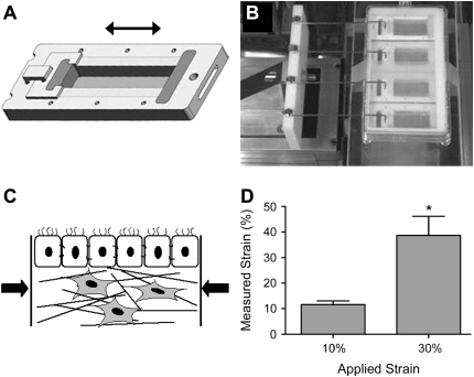
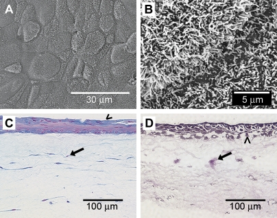
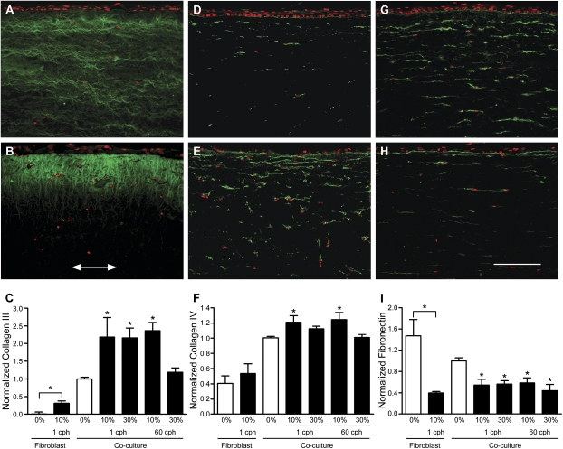
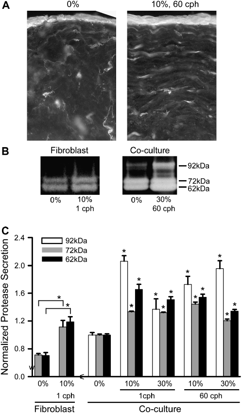
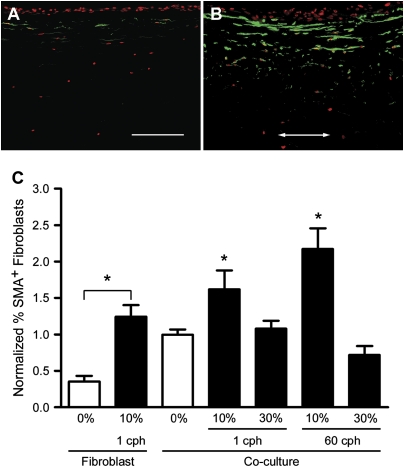
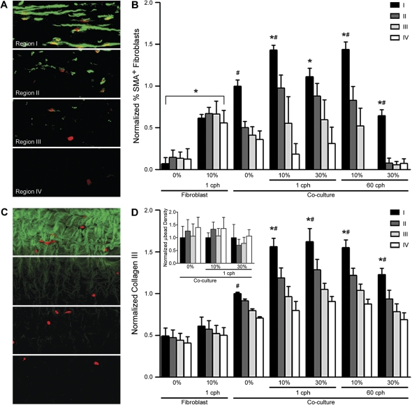
Airway wall remodeling is a hallmark of asthma, characterized by subepithelial thickening and extracellular matrix (ECM) remodeling. Mechanical stress due to hyperresponsive smooth muscle cells may contribute to this remodeling, but its relevance in a three-dimensional environment (where the ECM plays an important role in modulating stresses felt by cells) is unclear. To characterize the effects of dynamic compression in ECM remodeling in a physiologically relevant three-dimensional environment, a tissue-engineered human airway wall model with differentiated bronchial epithelial cells atop a collagen gel containing lung fibroblasts was used. Lateral compressive strain of 10 or 30% at 1 or 60 cycles per hour was applied using a novel straining device. ECM remodeling was assessed by immunohistochemistry and zymography. Dynamic strain, particularly at the lower magnitude, induced airway wall remodeling, as indicated by increased deposition of types III and IV collagen and increased secretion of matrix metalloproteinase-2 and -9. These changes paralleled increased myofibroblast differentiation and were fibroblast-dependent. Furthermore, the spatial pattern of type III collagen deposition correlated with that of myofibroblasts; both were concentrated near the epithelium and decreased diffusely away from the surface, indicating some epithelial control of the remodeling response. Thus, in a physiologically relevant three-dimensional model of the bronchial wall, dynamic compressive strain induced tissue remodeling that mimics many features of remodeling seen in asthma, in the absence of inflammation and dependent on epithelial-fibroblast signaling.


Figures

<b>Figure 1.</b>
Figure 1.
Dynamic strain model. (A) Individual wells with movable inner walls were designed to introduce lateral compressive strain to our three-dimensional human airway wall tissue models. (B) A computer-controlled motor-driven arm was coupled to the wells to impose the strain. (C) Lateral compression was introduced to the airway wall models. (D) The resulting strain, as measured by microbead displacement, was uniform throughout the length of the device, where applied strain of 10% and 30% at 60 cph translated to significantly different averages of 12% and 39% in gel, respectively (*P = 0.001); each was not significantly different from its applied strain of 10% and 30%.
<b>Figure 2.</b>
Figure 2.
Morphologic features of the three-dimensional human airway wall model. (A) SEM of the apical surface reveals a tightly packed epithelium. (B) Under higher magnification in SEM, a dense carpet of cilia is observed. (C) Alcian blue staining shows mucin production (blue) by epithelial cells (arrowhead); fibroblasts are uniformly distributed within the subepithelial tissue (arrow) with all nuclei in red. (D) Van Gieson and hematoxylin stains show fibroblasts (arrow) distributed throughout the collagen matrix beneath the epithelial layer (arrowhead).
<b>Figure 3.</b>
Figure 3.
Extracellular matrix protein deposition was altered by dynamic mechanical strain. All images show 10-μm paraffin-embedded sections, immunostained for types III collagen, IV collagen, and fibronectin (all in green), with a red nuclear counterstain; arrow indicates direction of strain and bar = 150 μm. Top row shows representative co-culture images from unstrained controls, while the middle row shows representative co-culture images from 10% strain, 1 cph. The bottom row shows the image intensity analysis averaged for all fibroblast-only and co-culture samples. (A) Type III collagen in unstrained versus (B) strained samples, where deposition was highest just beneath the epithelium and decreased with depth into the tissue (arrow indicates strain direction). (C) Type III collagen upregulation was greater for all strain levels except the highest strain rate applied (30% at 60 cph) in both fibroblast only and co-culture systems when compared with their respective static controls. (D) Type IV collagen expression in unstrained versus (E) strained samples, where deposition was also highest near the epithelium and decreased with depth. (F) Significant increase in type IV collagen was seen only with 10% strain in co-cultures. (G) Fibronectin production in unstrained versus (H) strained samples, where production was noticeably decreased. (I) Fibronectin quantification showed a significant decrease with all levels of strain and in the presence of the epithelium. *P < 0.05 compared with respective static controls.
<b>Figure 4.</b>
Figure 4.
Overall protease activity increased with all levels of strain. (A) In situ zymography on 25-μm cryosections show protease activity concentrated in the epithelium and around fibroblasts. (B) Representative gel zymograms from strained (10%, 1 cph) fibroblast-only versus co-culture models show bands at 62, 72, and 92 kD, corresponding to active MMP-2, pro–MMP-2, and pro–MMP-9, respectively. All were visibly increased with strain; in models with only fibroblasts, the 92-kD band was missing, indicating that the epithelial cells were critical in the release of pro–MMP-9. (C) Zymogram quantification demonstrates significant increases in proteolysis in all levels of strain normalized to their respective static controls. Static fibroblast-only samples showed no detectable MMP-9 and released ∼ 25% less MMP-2 than static co-cultures. *P < 0.05 compared with respective static controls.
<b>Figure 5.</b>
Figure 5.
Myofibroblast differentiation increased with 10% strain. Representative (A) static and (B) strained (10%, 1 cph) sections from co-cultures show the upregulation of α-SMA+ cells (green with red nuclear counterstain), particularly close to the epithelium, by dynamic strain. Arrow indicates direction of strain (bar = 150 μm). (C) Quantification of α-SMA+ cells demonstrates that 10% strain caused significant increases in α-SMA expression compared with respective static controls; cell differentiation was increased by 62% at 1 cph and by 117% at the higher frequency of 60 cph. *P < 0.05 compared with respective static controls.
<b>Figure 6.</b>
Figure 6.
The spatial distribution of myofibroblasts generally correlated with the spatial distribution of type III collagen deposition and was concentrated close to the basal side of the epithelium. (A) Immunostained thin sections were divided into four equal regions for intensity analysis as shown for α-SMA; each region had a depth of 88 μm. (B) Myofibroblasts, as detected by α-SMA expression, were found in greater numbers in region I, and diminished in regions farther away from the epithelium. Furthermore, only 10% strain resulted in significantly greater myofibroblast expression compared with region I in static control co-cultures. In fibroblast-only cultures, 10% strain resulted in a uniform increase of α-SMA expression. (C) Likewise, thin sections were immunostained for type III collagen and divided into equal regions. (D) Type III collagen production was also significantly increased in region I near the epithelium under all conditions, and all strain conditions resulted in significantly greater production in region I as compared with static controls for co-cultures. Regional difference in type III collagen production was not observed in fibroblast-only cultures. The inset bar graph shows uniform distribution of microbeads throughout various depths in co-cultures in the presence and absence of strain. *P < 0.01 compared with respective static controls and #P < 0.05 compared with respective regions II–IV.

Similar articles

Cited by

References

    1. James A. Airway remodeling in asthma. Curr Opin Pulm Med 2005;11: 1–6. - PubMed
    1. Homer RJ, Elias JA. Airway remodeling in asthma: therapeutic implications of mechanisms. Physiology (Bethesda) 2005;20:28–35. - PubMed
    1. Cohn L, Elias JA, Chupp GL. Asthma: mechanisms of disease persistence and progression. Annu Rev Immunol 2004;22:789–815. - PubMed
    1. McParland BE, Macklem PT, Pare PD. Airway wall remodeling: friend or foe? J Appl Physiol 2003;95:426–434. - PubMed
    1. Jeffery PK. Remodeling in asthma and chronic obstructive lung disease. Am J Respir Crit Care Med 2001;164:S28–S38. - PubMed

Publication types

Substances