. 2006 Sep;35(3):314-9.
doi: 10.1165/rcmb.2005-0428OC. Epub 2006 Apr 6.

Cigarette smoke-induced Egr-1 upregulates proinflammatory cytokines in pulmonary epithelial cells

Affiliations

Cigarette smoke-induced Egr-1 upregulates proinflammatory cytokines in pulmonary epithelial cells

Paul R Reynolds et al. Am J Respir Cell Mol Biol. 2006 Sep.

Abstract

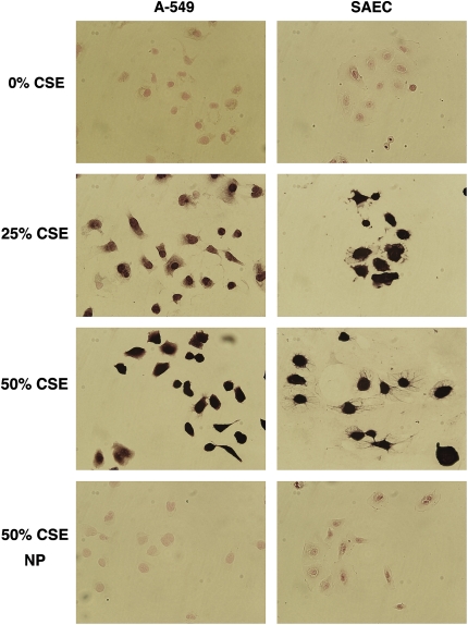
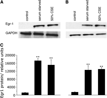
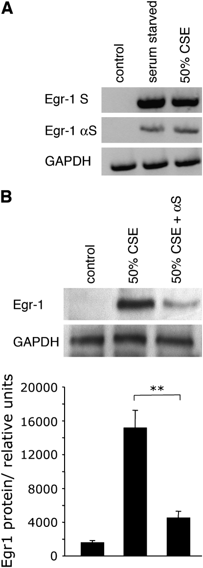
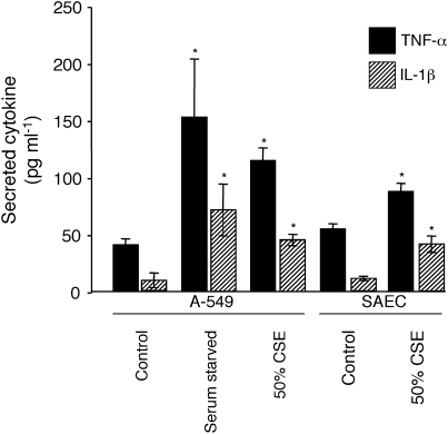
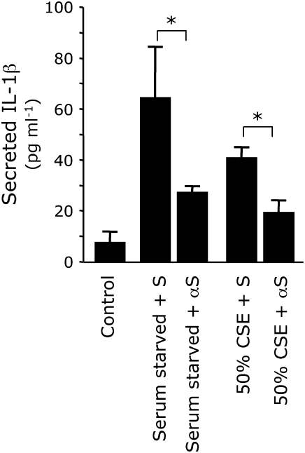
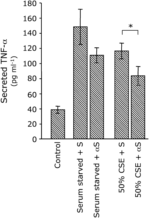
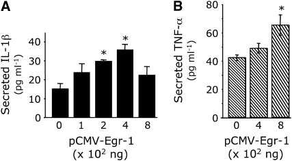
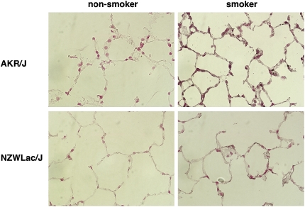
Chronic obstructive pulmonary disease (COPD) is the fourth leading cause of death worldwide and is a progressive and irreversible disorder. Cigarette smoking is associated with 80-90% of COPD cases; however, the genes involved in COPD-associated emphysema and chronic inflammation are poorly understood. It was recently demonstrated that early growth response gene 1 (Egr-1) is significantly upregulated in the lungs of smokers with COPD (Ning W and coworkers, Proc Natl Acad Sci 2004;101:14895-14900). We hypothesized that Egr-1 is activated in pulmonary epithelial cells during exposure to cigarette smoke extract (CSE). Using immunohistochemistry, we demonstrated that pulmonary adenocarcinoma cells (A-549) and primary epithelial cells lacking basal Egr-1 markedly induce Egr-1 expression after CSE exposure. To evaluate Egr-1-specific effects, we used antisense (alphaS) oligodeoxynucleotides (ODN) to knock down Egr-1 expression. Incorporation of Egr-1 alphaS ODN significantly decreased CSE-induced Egr-1 mRNA and protein, while sense ODN had no effect. Via Egr-1-mediated mechanisms, IL-1beta and TNF-alpha were significantly upregulated in pulmonary epithelial cells exposed to CSE or transfected with Egr-1. To investigate the relationship between Egr-1 induction by smoking and susceptibility to emphysema, we determined Egr-1 expression in strains of mice with different susceptibilities for the development of smoking-induced emphysema. Egr-1 was markedly increased in the lungs of emphysema-susceptible AKR/J mice chronically exposed to cigarette smoke, but only minimally increased in resistant NZWLac/J mice. In conclusion, Egr-1 is induced by cigarette smoke and functions in proinflammatory mechanisms that likely contribute to the development of COPD in the lungs of smokers.


Figures

<b>Figure 1.</b>
Figure 1.
Exposure of A-549 cells and SAECs to CSE upregulates Egr-1 expression. Cells at 40–50% confluence were incubated in media alone, 25% CSE, or 50% CSE for 2 h, immediately fixed with 4% paraformaldehyde, and immunostained for Egr-1 protein expression. Egr-1 expression was not detected in control cells grown in medium alone. Egr-1 was markedly induced in cells exposed to 25% and 50% CSE. Experiments without primary antibodies revealed no immunoreactivity after exposure to 50% CSE. Images are at ×80 magnification.
<b>Figure 2.</b>
Figure 2.
CSE induces Egr-1 expression in human pulmonary adenocarcinoma cells (A-549) and Primary SAECs. (A and B) Representative Western blot analysis reveals no Egr-1 expression in cells grown in medium alone (control). Significant upregulation of Egr-1 was observed in serum-starved cells and in cells exposed to 50% CSE for 2 h. (C) Relative densitometry was quantified by using NIH Image-J software. Significant differences in Egr-1 protein expression as compared with control are noted at P ⩽ 0.01 (**).
<b>Figure 3.</b>
Figure 3.
Egr-1 αS ODNs inhibit Egr-1 mRNA and protein expression. (A) A-549 cells were transfected with Egr-1 sense (S) or antisense (αS) oligonucleotides (10 μM) and serum starved or exposed to CSE 12 h after transfection for 2 h. Total RNA was isolated, 1.0 μg was reverse transcribed, and the resulting cDNA was subjected to PCR analysis. αS oligonucleotides significantly decreased Egr-1 mRNA expression. (B) Cells manipulated as outlined above were lysed and subjected to Western blot analysis. Significant αS ODN-mediated decreases in Egr-1 expression were detected after serum starvation or exposure to CSE. Relative densitometry was quantified by using NIH Image-J software. Significant differences in Egr-1 protein expression are noted at P ⩽ 0.05 (*) and P ⩽ 0.01 (**).
<b>Figure 4.</b>
Figure 4.
CSE exposure induces proinflammatory cytokine secretion. A-549 cells were serum starved or exposed to 50% CSE for 2 h and medium was immediately assayed for secreted IL-1β (shaded bars) and TNF-α (solid bars) by ELISA. Compared with basal levels observed from cells grown in DMEM-10% FCS (control), cells exposed to media lacking serum or 50% CSE had significantly elevated levels of secreted IL-1β and TNF-α. SAECs exposed to 50% CSE also induced cytokine secretion. Data presented are from two experiments, each performed in triplicate. *P ⩽ 0.05.
<b>Figure 5.</b>
Figure 5.
Egr-1 αS ODNs inhibit CSE-induced IL-1β secretion. A-549 cells were transfected with Egr-1 sense (S) or antisense (αS) oligonucleotides (10 μM) and serum starved or exposed to CSE 12 h after transfection for 2 h. Medium was isolated and subjected to ELISA. Secreted IL-1β was significantly decreased by Egr-1 αS oligonucleotides after serum starvation or CSE exposure. Data presented are from two experiments, each performed in triplicate. *P ⩽ 0.05.
<b>Figure 6.</b>
Figure 6.
Egr-1 αS ODNs inhibit CSE-induced TNF-α secretion. A-549 cells were transfected with Egr-1 sense (S) or antisense (αS) oligonucleotides (10 μM) and serum starved or exposed to CSE 12 h after transfection for 2 h. Medium was isolated and subjected to ELISA. Secreted TNF-α was not significantly decreased by Egr-1 αS ODNs after serum starvation. TNF-α secretion was significantly inhibited by Egr-1 αS ODNs in cells exposed to 50% CSE. Data presented are from two experiments, each performed in triplicate. *P ⩽ 0.05.
<b>Figure 7.</b>
Figure 7.
Exogenous Egr-1 induces proinflammatory cytokine secretion. In the absence of CSE, A-549 cells were transfected with Egr-1 (pCMV-Egr-1), and secreted IL-1β or TNF-α in the medium was assessed by ELISA. While transfection experiments with 200–400 ng Egr-1 significantly induced IL-1β secretion, TNF-α secretion was significantly elevated only after 800 ng of Egr-1 was transfected. Data presented are from two experiments, each performed in triplicate. *P ⩽ 0.05.
<b>Figure 8.</b>
Figure 8.
Egr-1 is induced in the lungs of mice exposed to cigarette smoke. Cigarette smoke–induced emphysema hypersensitive (AKR/J) and resistant (NZWLac/J) mice were maintained in environments either lacking (nonsmokers) or including cigarette smoke (smokers) for 6 mo. Immunohistochemistry reveals minimal Egr-1 expression in nonsmoker lungs of both strains; however Egr-1 expression was markedly increased in AKR/J smoker lungs. Egr-1 was minimally increased in the lungs of smoker NZWLac/J mice resistant to cigarette smoke–induced emphysema. Control sections incubated in serum alone had no immunoreactivity (not shown). Figures are at ×100 magnification.

Similar articles

Cited by

References

    1. Voelkel NF, MacNee W. Chronic obstructive lung diseases. Hamilton, ON: B. C. Decker; 2002. pp. 90–113.
    1. Petty TL. COPD in perspective. Chest 2002;121:116S–120S. - PubMed
    1. Sethi JM, Rochester CL. Smoking and chronic obstructive pulmonary disease. Clin Chest Med 2000;21:67–86. - PubMed
    1. Mayer AS, Newman LS. Genetic and environmental modulation of chronic obstructive pulmonary disease. Respir Physiol 2001;128:3–11. - PubMed
    1. Ning W, Li CJ, Kaminski N, Feghali-Bostwick CA, Alber SM, Di YP, Otterbein SL, Song R, Hayashi S, Zhou Z, et al. Comprehensive gene expression profiles reveal pathways related to the pathogenesis of chronic obstructive pulmonary disease. Proc Natl Acad Sci USA 2004;101:14895–14900. - PMC - PubMed

Publication types